When > 10, consider autosomal dominant polycystic liver disease (ADPLD) or biliary hamartomas
• Sharply defined margins, thin walls
• Water density (-10 to +10 HU)
• Usually no or few thin septations
No mural nodularity or wall calcification
• Hemorrhage into cyst may simulate tumor
No enhancement of “solid” material
Varied MR signal intensity (due to mixed blood products)
• US: Anechoic mass, accentuated through transmission
Smooth borders; thin or invisible wall
• Size varies from few mm to > 20 cm
Rarely are the cysts of similar size
Helps to differentiate from biliary hamartomas, which are all usually < 15 mm
TOP DIFFERENTIAL DIAGNOSES
• AD polycystic disease, liver
• Cystic or necrotic metastases
• Biliary cystadenocarcinoma
• Biliary hamartomas
• Ciliated hepatic foregut cyst
• Hepatic cavernous hemangioma
• Biloma
• Hepatic pyogenic abscess
• Hydatid (echinococcal) disease
DIAGNOSTIC CHECKLIST
• Sonography shows cyst morphology better than CT
(Left) Axial CECT shows a spherical hepatic mass with water density and homogeneous contents. No internal debris or wall irregularities are present. This is a classic simple cyst.
(Right) Ultrasound in the same patient shows an anechoic mass with accentuated through-transmission . Either CT or US would have been sufficient to establish the diagnosis in this patient.
(Left) Axial T1WI MR shows a large, cystic, hepatic mass that has homogeneous low intensity and several thin septa .
(Right) Coronal T2WI shows uniform high intensity and septa . The cyst has remained stable for years, and no other evaluation or intervention was performed.
TERMINOLOGY
Synonyms
• Simple hepatic or bile duct cyst
Definitions
• Benign, congenital, developmental lesion derived from biliary endothelium
IMAGING
General Features
• Best diagnostic clue
Anechoic lesion with increased through-transmission and no mural nodularity on US
• Location
Any location within liver
• Size
Varies from few mm to > 20 cm
– Rarely are the cysts of similar size
– Helps to differentiate from biliary hamartomas, which are all usually < 15 mm
• Morphology
Spherical or oval, well marginated
• Key concepts
Classified based on etiology and pathogenesis
Congenital or developmental: Simple hepatic or bile duct cyst
– Often multiple: Usually < 10
– No communication with bile ducts
When > 10 in number, fibropolycystic disease must be considered


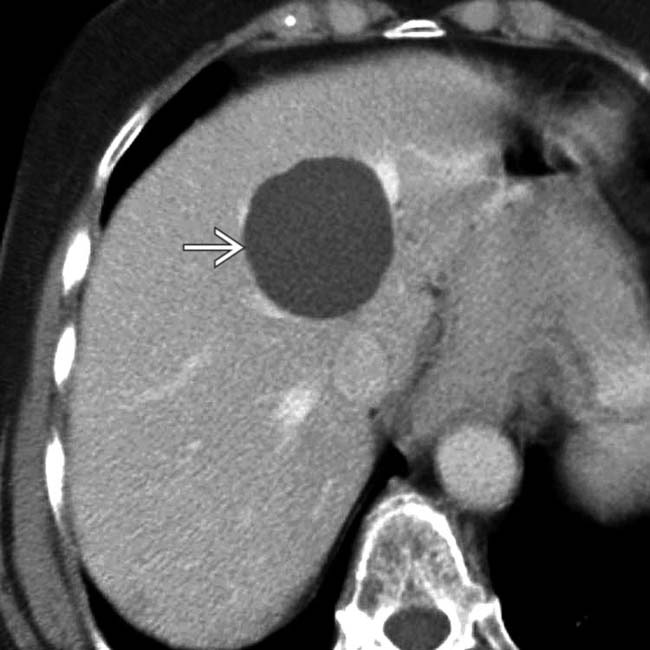
 with water density and homogeneous contents. No internal debris or wall irregularities are present. This is a classic simple cyst.
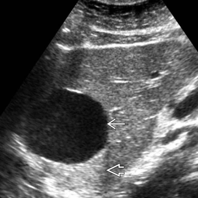
 with accentuated through-transmission
  . Either CT or US would have been sufficient to establish the diagnosis in this patient.
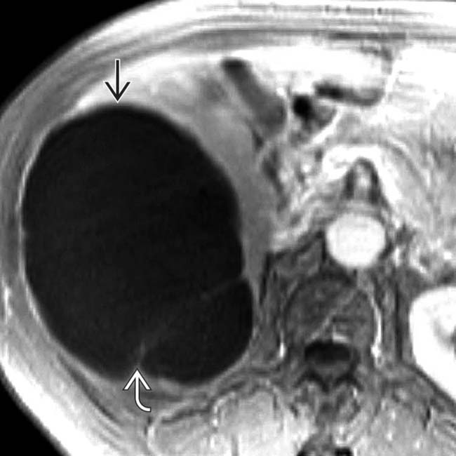
 that has homogeneous low intensity and several thin septa
  .
.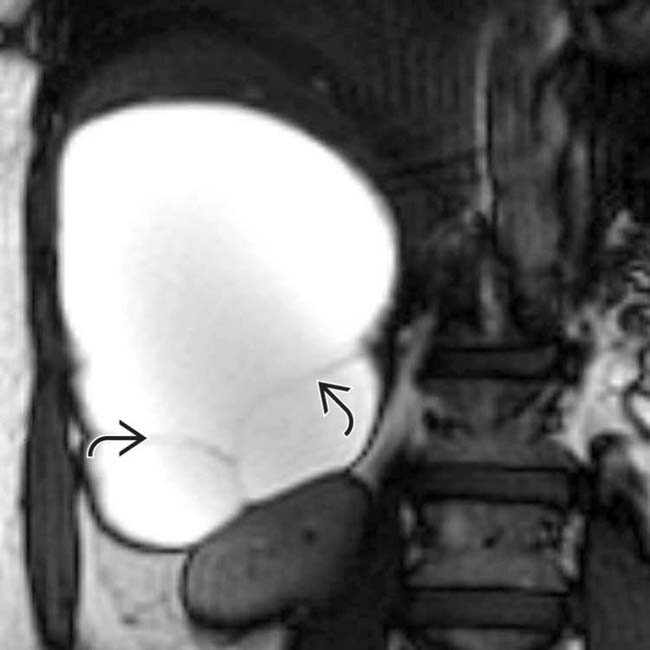
 . The cyst has remained stable for years, and no other evaluation or intervention was performed.


