Contains multiple internal “daughter” cysts of lower density than “mother” cyst (exocyst)
Curvilinear ring-like calcification of pericyst (wall)
Calcified wall: Usually indicates no active infection if completely circumferential
Dilated intrahepatic bile duct: Due to compression or rupture of cyst into bile ducts
US: Multiseptate cyst with “daughter” cysts and echogenic material between cysts
Water lily sign: Cyst with floating, undulating membrane and detached endocyst
• Echinococcus multilocularis (alveolaris): Less common but aggressive, tumor-like form
Extensive, infiltrative cystic and solid masses of low density (14-40 HU)
Margins are irregular and ill defined
Simulates primary or secondary malignant tumor
TOP DIFFERENTIAL DIAGNOSES
• Biliary cystadenocarcinoma
Rare, solitary, multiseptate, water density cystic mass
• Hepatic pyogenic abscess
“Cluster of grapes”: Confluent complex cystic lesions
• “Cystic” metastases
• Hemorrhagic or infected cyst
CLINICAL ISSUES
• Cysts: Initially asymptomatic
• Symptomatic with ↑ in size or cyst rupture
Rupture into biliary tree, peritoneal or pleural cavity is not rare
(Left) Graphic shows a hydatid cyst within the liver with a peripheral fibrous capsule (pericyst) and numerous “daughter” cysts within.
(Right) Gross photograph of liver shows a hydatid cyst containing multiple “daughter” cysts . The fibrous rim or pericyst can be seen surrounding the cyst. (Courtesy K. Caradine, MD.)
(Left) This 29-year-old woman emigrated to the USA from Jordan. Axial CT shows a classic spherical mass, exophytic from the left lobe of the liver. Note the thick, fibrotic wall (pericyst) and the presence of peripheral “daughter” cysts within the larger cyst.
(Right) A coronal-reformatted CT image from the same patient clearly demonstrates the “daughter” cysts within the larger “mother” cyst (exocyst). A similar lesion was present within the left hepatic lobe (not shown).
TERMINOLOGY
Synonyms
• Echinococcal or hydatid disease
• Echinococcosis
Definitions
• Infection of humans caused by larval stage of Echinococcus species
IMAGING
General Features
• Best diagnostic clue
Large, well-defined, cystic liver mass with numerous peripheral “daughter” cysts
• Size
Average size: 5 cm
Maximum size: Up to 50 cm
May contain up to 1.5 liters of fluid
• Key concepts
Echinococcus granulosus: Most common cause of hydatid disease
– Up to 60% of cysts are multiple
Echinococcus multilocularis (alveolaris): Less common but aggressive, tumor-like form
Radiographic Findings
• Radiography
E. granulosus
– Curvilinear or ring-like pericyst calcification
– Seen on abdominal plain films in ∼ 20-30% of affected patients
E. multilocularis (alveolaris)
– Microcalcifications in 50% of cases
• ERCP
Hydatid cyst may communicate with biliary tree
– Gallbladder much less common
CT Findings
• CECT
E. granulosus
– Uni- or multilocular, well-defined cysts
– Contain multiple peripheral “daughter” cysts of lower density than “mother” cyst
– Curvilinear ring-like calcification of pericyst (wall)
Usually indicates no active infection if completely circumferential
– Enhancement of cyst wall and septa
– Dilated intrahepatic bile duct (IHBD)
Due to compression or rupture of cyst into ducts
E. multilocularis (alveolaris)
– Extensive, infiltrative cystic and solid masses of low density (14-40 HU)
– Margins are irregular and ill defined
– Amorphous type of calcification
– Simulates primary or secondary malignant tumor
– Minimal enhancement of noncalcified portions
MR Findings
• T1WI
Rim (pericyst): Hypointense (fibrous component)
“Mother” cyst (hydatid matrix)
– Usually intermediate signal intensity
– Rarely hyperintense
“Daughter” cysts: Less signal intensity than “mother” cyst (matrix)
Floating membrane: Low signal intensity
Calcifications: Difficult to identify on MR images
– Display low signal on both T1WI & T2WI
• T2WI
Rim (pericyst): Hypointense (fibrous component)
1st echo T2WI: Increased signal intensity
– “Mother” cysts more than “daughter” cysts
Strong T2WI: Hyperintense
– “Mother” and “daughter” cysts have same intensity
Floating membrane
– Low to intermediate signal intensity
• T1WI C+
E. granulosus
– Enhancement of cyst wall and septations
Only gold members can continue reading. Log In or Register to continue


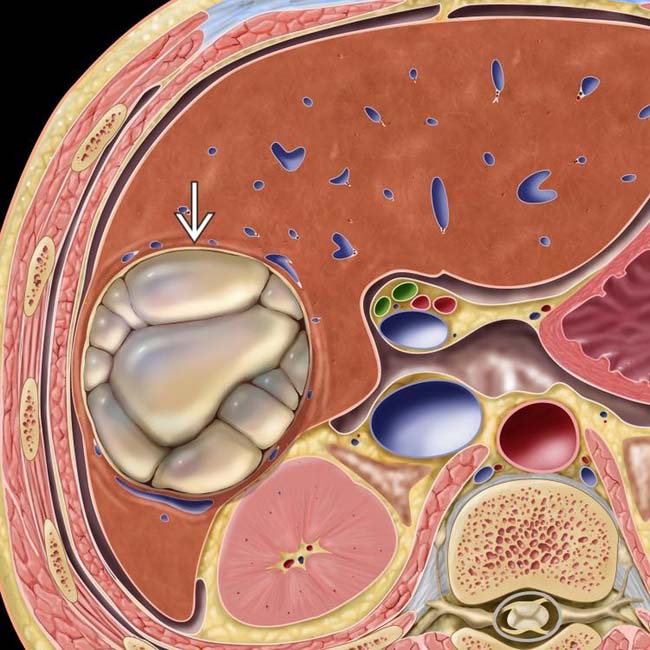
 within the liver with a peripheral fibrous capsule (pericyst) and numerous “daughter” cysts within.
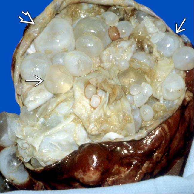
 . The fibrous rim
  or pericyst can be seen surrounding the cyst. (Courtesy K. Caradine, MD.)
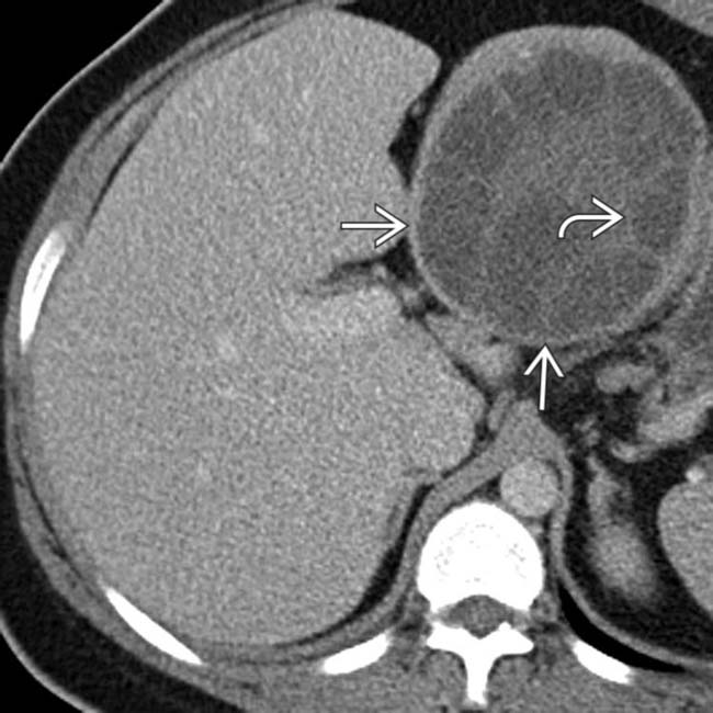
 and the presence of peripheral “daughter” cysts
  within the larger cyst.
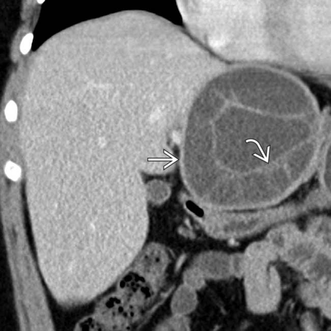
 within the larger “mother” cyst
  (exocyst). A similar lesion was present within the left hepatic lobe (not shown).


 E. granulosus


