Metastases: Hypointense lesions made more apparent compared with bright enhancement of liver on delayed phase imaging
• CECT is usually best as “whole body” screening test
Even better if combined as PET/CT
Metastases and lymphoma are usually FDG-avid masses within liver
• Decision for thermal ablation or surgical resection
May require most sensitive tests (gadoxetate-enhanced MR, PET/CT, or intraoperative US)
TOP DIFFERENTIAL DIAGNOSES
• Multifocal fatty infiltration (steatosis)
• Multiple benign masses
• Multifocal hepatocellular carcinoma or cholangiocarcinoma
DIAGNOSTIC CHECKLIST
• In absence of a known primary tumor or other metastases:
Hepatic lesions that are “too small to characterize” rarely represent metastases
Lesions that are lower than blood density on NECT rarely represent metastases
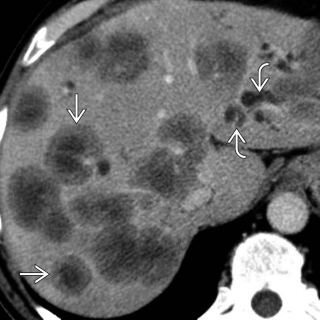
(Left) Axial CECT shows multiple spherical liver lesions with a “target” appearance. This is the most typical appearance for liver metastases, especially from colon cancer. Also note the focally dilated bile ducts due to compression by the metastases.
(Right) Color Doppler ultrasound in the same patient shows multiple spherical liver lesions with a “target” appearance , some containing visible blood vessels . This is the typical appearance of metastatic colorectal carcinoma.
(Left) Axial T1WI C+ MR in a patient with metastatic colon cancer shows multiple liver metastases with several typical features, including a continuous ring of enhancement .
(Right) Axial T2WI FS MR in the same patient shows heterogeneous hyperintensity within the hepatic metastases . Most metastases are heterogeneously hyperintense on T2WI and hypovascular and hypointense on T1WI.
TERMINOLOGY
Abbreviations
•
Synonyms
Definitions
• Lymphoma: Neoplasm of lymphoid tissues
• Metastases: Malignant spread of neoplasm to hepatic parenchyma


 with a “target” appearance. This is the most typical appearance for liver metastases, especially from colon cancer. Also note the focally dilated bile ducts
  due to compression by the metastases.
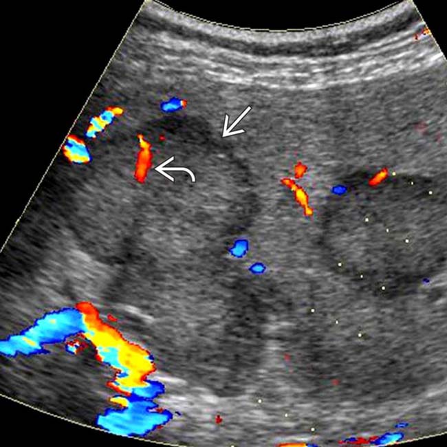
 , some containing visible blood vessels
  . This is the typical appearance of metastatic colorectal carcinoma.
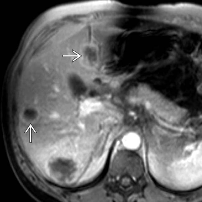
 .
.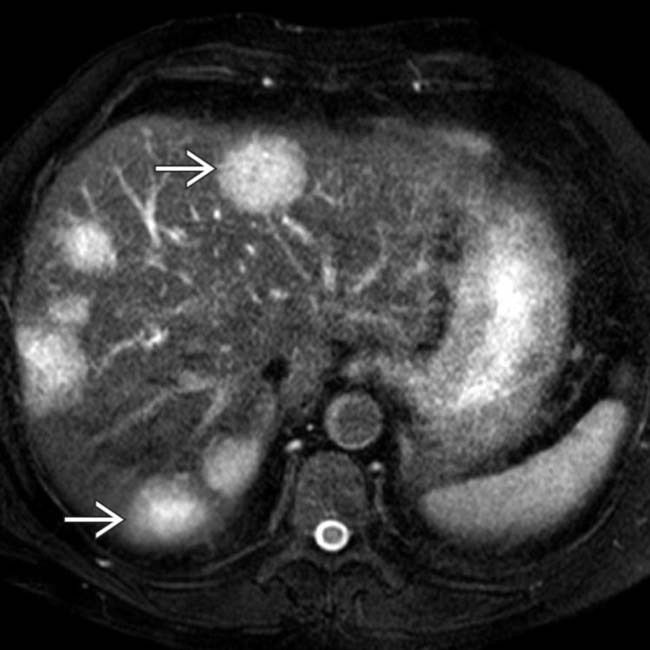
 . Most metastases are heterogeneously hyperintense on T2WI and hypovascular and hypointense on T1WI.


 Bull’s-eye or “target” metastatic lesions


