Capsular calcification (parallel or perpendicular to liver surface)
• Portal hypertension in advanced disease
Splenomegaly and varices
• US: Bull’s-eye lesion: Represents anechoic portal vein surrounded by echogenic mantle of fibrous tissue
Hyperechoic and thickened walls of portal venules
Network of echogenic septa outlining polygonal areas of normal-appearing liver
• US elastography demonstrates hepatic fibrosis
• MR shows same morphologic signs as CT of liver damage and portal hypertension
MR elastography provides a measure of extent of hepatic fibrosis, which may determine therapy and prognosis
TOP DIFFERENTIAL DIAGNOSES
• Hepatic cirrhosis
Often has widened fissures but not as much periportal fibrosis or calcification as with schistosomiasis
CLINICAL ISSUES
• Most common cause of hepatic fibrosis in the world
Over 200,000,000 persons, mostly in tropics
• Different Schistosoma species affect urinary tract more than liver
• Oral praziquantel for treatment
DIAGNOSTIC CHECKLIST
• Exclude other causes of hepatic fibrosis or cirrhosis
• Hepatic mosaic “tortoise shell” pattern of fibrosis and calcification
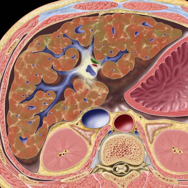
(Left) Graphic shows striking periportal edema and fibrosis with widened fissures between hepatic segments.
(Right) Axial CT shows signs of portal hypertension, including large varices and splenomegaly. Note the extraordinarily widened hepatic fissures deeply dividing the segments of the liver along the portal vein branches. This is a characteristic feature of hepatic schistosomiasis; the appearance of the liver has been described as that of a tortoise shell.
(Left) In low-power micropathology, portal tracts show chronic inflammation, luminal narrowing, and several granulomas containing degenerated Schistosoma ova . (Courtesy J. Misdraji, MD.)
(Right) Axial NECT of the liver shows extensive calcification and peripheral fibrosis in patterns such as thin curvilinear , subcapsular band-like , and confluent . The predominantly peripheral location and calcification of the fibrotic regions are distinguishing features from viral or alcoholic cirrhosis.
TERMINOLOGY
Synonyms
• Bilharzia, bilharziasis, blood fluke
Definitions
• Hepatic parasitic infestation by Schistosoma species
IMAGING
General Features
• Best diagnostic clue
Periportal fibrotic bands and widened fissures with calcification
• Location
Diffuse throughout liver
• Morphology
Distortion of liver architecture and surface contour by extension of periportal fibrosis
CT Findings
• CECT
Hepatic involvement
– “Tortoise shell” or “turtle back” appearance
Represents calcified septa, aligned along and perpendicular to liver capsule
– Capsular calcification
– Markedly dysmorphic liver with peripheral atrophy, caudate hypertrophy
– Periportal edema, fibrosis, volume loss
– Splenomegaly and varices
Colonic involvement
– Ulceration of mucosa
– Submucosal edema + fibrosis
– May progress to calcification of colonic wall
MR Findings
• Shows same morphologic signs as CT of liver damage and portal hypertension
• MR elastography provides a measure of extent of hepatic fibrosis, which may determine therapy and prognosis
Ultrasonographic Findings
• Grayscale ultrasound
Hepatomegaly in early stages
Atrophic liver in late stage (fibrosis and portal hypertension)
Irregular/notched liver surface
Echogenic granulomata
– Peripheral/subcapsular location
– Egg deposited in terminal portal venule, resulting in inflammatory reaction
Periportal fibrosis
– Most severe at porta hepatis
– Widened portal tracts
– Hyperechoic & thickened walls of portal veins
– Described as “clay-pipestem” fibrosis
– Bull’s-eye lesion: Represents anechoic portal vein surrounded by echogenic mantle of fibrous tissue
Mosaic pattern
– Network of echogenic septa outlining polygonal areas of normal-appearing liver
– Represents complete septal fibrosis (inflammation and fibrosis as reaction to embolized eggs)


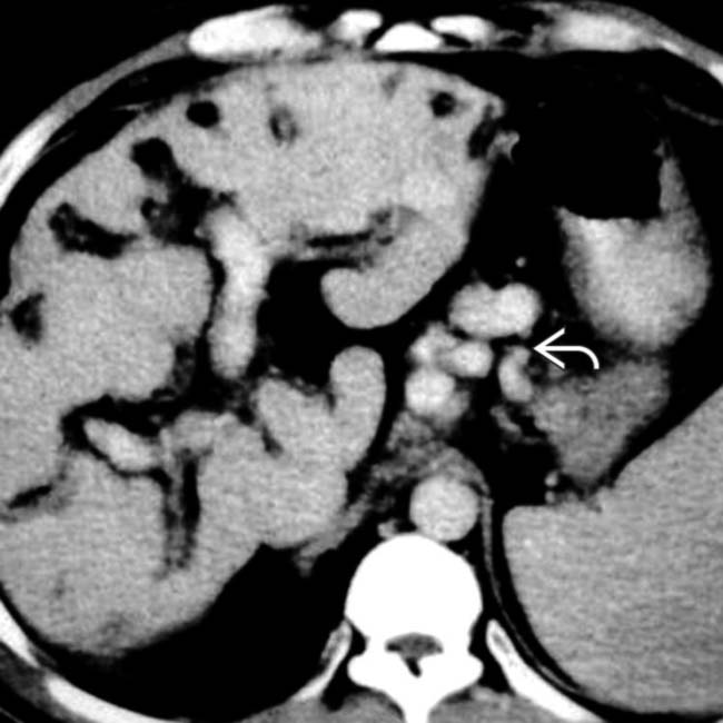
 and splenomegaly. Note the extraordinarily widened hepatic fissures deeply dividing the segments of the liver along the portal vein branches. This is a characteristic feature of hepatic schistosomiasis; the appearance of the liver has been described as that of a tortoise shell.
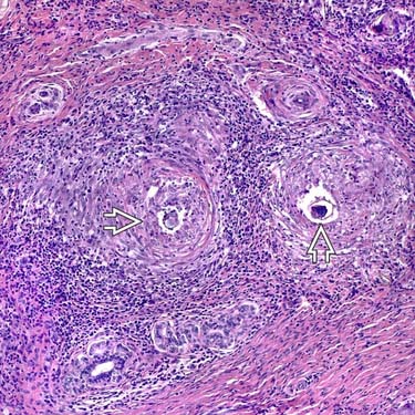
 . (Courtesy J. Misdraji, MD.)
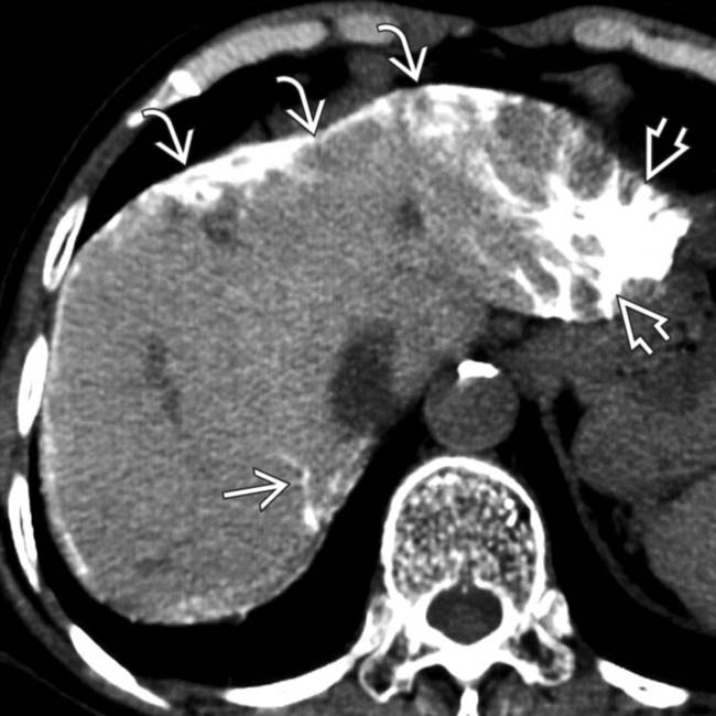
 , subcapsular band-like
  , and confluent
  . The predominantly peripheral location and calcification of the fibrotic regions are distinguishing features from viral or alcoholic cirrhosis.


