Gadolinium enhancement required to detect very small lesions
TOP DIFFERENTIAL DIAGNOSES
• Metastases
• Lymphomatous/leukemic foci in liver
• Biliary hamartomas
• Caroli disease
PATHOLOGY
• Originates from intestinal seeding of portal venous circulation
• Candida albicans: Most common cause of fungal microabscesses
CLINICAL ISSUES
• Most common signs/symptoms
Asymptomatic or abdominal pain and fever
Erythematous papules on skin
• Clinical profile: Immunocompromised patient recovering from neutropenia
High incidence in transplant patients with fungal colonization
DIAGNOSTIC CHECKLIST
• Rule out other innumerable hypodense liver lesions
• Biopsy specimen for histology/microbiology
(Left) High-power view of hepatic miliary tuberculosis shows a granuloma with focal eosinophilic granular necrotic material and several multinucleated giant cells . (Courtesy J. Misdraji, MD.)
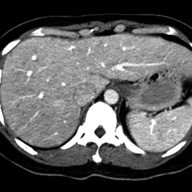
(Right) Axial CECT of a woman with breast cancer, fever, and liver dysfunction demonstrates multiple small, low-density lesions due to hepatic candidiasis. Metastases would uncommonly be so numerous and small.
(Left) Axial CECT in a patient undergoing chemotherapy for acute leukemia, now presenting with a fever, demonstrates fungal abscesses due to Candida. Note the multiple small (< 1 cm) lesions in all lobes of the liver.
(Right) Axial CECT of a 33-year-old woman with non-Hodgkin lymphoma presenting with a recent spike in temperature shows innumerable tiny hypodense lesions in the liver and spleen due to candidiasis. Lymphomatous parenchymal involvement is rarely detected as such small discrete lesions.
TERMINOLOGY
Definitions
• Opportunistic infection of liver (± other viscera), usually by fungal or mycobacterial organisms
IMAGING
General Features
• Best diagnostic clue
Multiple well-defined, rounded microabscesses in liver
• Location
May involve multiple organs
– Liver, spleen, kidneys, lungs
• Size
Microabscesses < 2 cm
• Morphology
Spherical with complex fluid center, ± enhancing capsule
CT Findings
• NECT
Multiple small, hypodense lesions
± scattered areas of calcific density (healing phase)
• CECT
Biphasic CT may be more accurate than venous phase only
– Nonenhancing hypodense centers
– ± peripheral rim enhancement
Central or eccentric “dot” felt to represent hyphae
Rarely may demonstrate unusual central enhancement on arterial phase with double peripheral rim


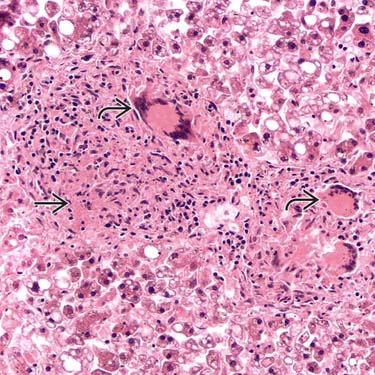
 and several multinucleated giant cells
  . (Courtesy J. Misdraji, MD.)
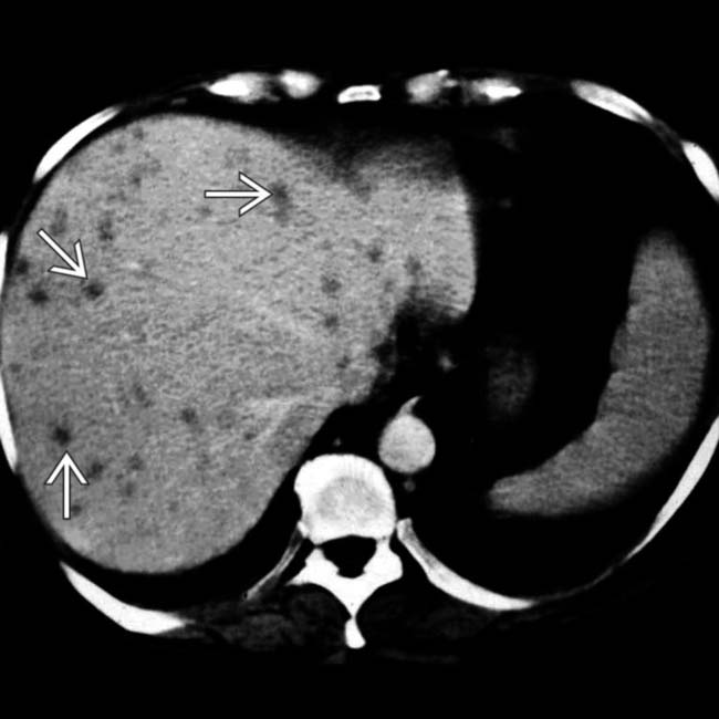
 due to hepatic candidiasis. Metastases would uncommonly be so numerous and small.
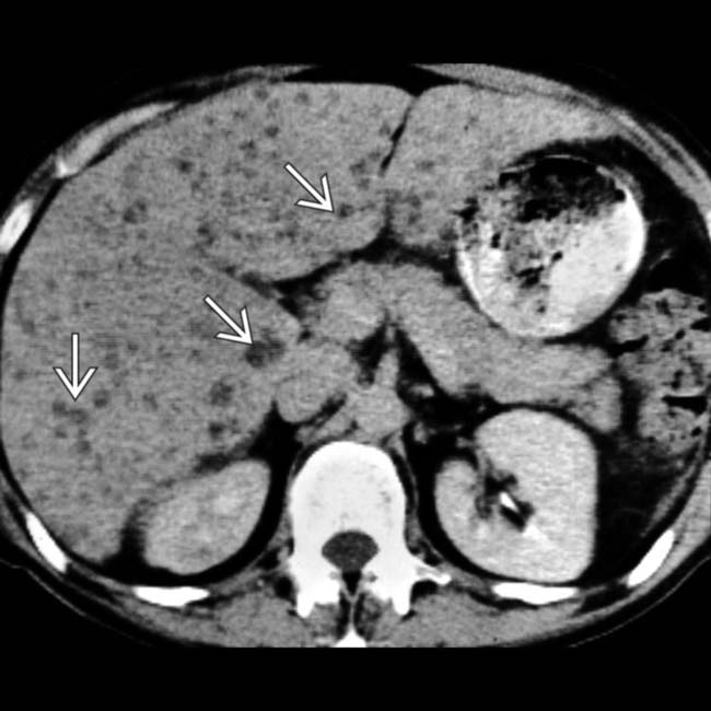
 in all lobes of the liver.