Fig. 1
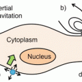
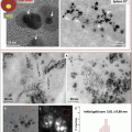
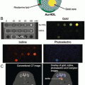
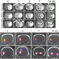
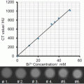
Comparison of the capability of different large animal and human molecular imaging modalities. The matrix uses a log-log scale to indicate sensitivity on the vertical axis and spatial resolution on the horizontal axis. Maximum resolution corresponds to the left limit on the horizontal axis and maximum sample diameter corresponds to the right limit. The sensitivity range for PET is taken from Meikle et al. [71] and spatial resolution from Moses [6]. The sensitivity for SPECT is also taken from Meikle et al. [71] and spatial resolution is estimated at 1.2 cm; however, this is dependent on collimator choice and distance from collimator to object [72]. For fluorescence and bioluminescence imaging, resolution limit can be higher than the optical wavelength [73], i.e., much better than 100 μm, but decreases very quickly for an object of any thickness. A 1 cm limit for thickness is generous and corresponds to hybrid methods such as photoacoustic imaging [74]. For MRI with paramagnetic contrast agents, 1 mm3 resolution at 3 T is easily achievable, with sample size being limited by available magnet bore [75]. For MRI with iron particles, refer to the section on estimated limits of sensitivity of a MRI reporter gene. For magnetic resonance spectroscopy (MRS), the spatial resolution for 31P, 19F, 23Na, or 1H (from non-water protons) is limited to approximately 1 cm due to gyromagnetic ratio and/or concentration of the isotope [76]. The values for CT were taken from Gore et al. [76]
The idea of using magnetotactic bacterial genes as noninvasive reporters of cellular activity for molecular MRI has recently been put forward [8–10]. Magnetotactic bacteria form magnetosomes [11, 12], membrane-enclosed iron biominerals that respond to the earth’s magnetic field and enable magnetotaxis. With these attributes, the motile microaerophilic bacteria may navigate toward their preferred oxic-anoxic zones in aquatic sediments [13]. Magnetosomes are also similar in size and magnetic properties [14] to superparamagnetic iron oxide (SPIO) nanoparticles. While the latter have been used successfully to track cells in both research and clinical settings [15–18], within the genetic determinants of magnetosome synthesis is an opportunity to specify MR contrast as a direct response of select gene expression.
Approximately 20 years ago, a few reports were published about genes related to the magnetic properties of species of Alphaproteobacteria [19, 20]; although, it is only in the last decade that a clearer definition of the magnetosome and its constituent proteins has emerged [21]. The magnetosome is formed by a group of nonessential genes, suggesting that this bacterial structure is dispensable and confers an auxiliary function to the cell, i.e., magnetotaxis . When present, the magnetosome comprises an iron biomineral that is compartmentalized within a specialized lipid bilayer, protecting the cell from iron toxicity and confining the biomineral to a defined subcellular location. As detailed below, the magnetosome membrane contains a number of proteins that direct its location and crystal composition, size, and shape. In short, the magnetosome is an ideal structure by which cellular and molecular MRI may be refined.
Recent progress in defining the magnetosome in molecular terms provides an opportunity to further develop genetically engineered, MR contrast for effective molecular MRI [22, 23]. Such a tool would address the critical need to identify molecular activities that define the early stages of disease progression, ahead of the irreversible damage to tissue that leads to chronic illness . This is where the true strength of noninvasive reporter gene expression lies. The ability to detect transcription factor activity that prompts disease-related changes in gene expression is the key to understanding many, if not most medical conditions , including cancer [24, 25], inflammation [26, 27], and the fibrosis that leads to heart disease [28, 29]. Effective use of MRI reporter gene expression vectors, which create and strictly regulate magnetosome-like particles in mammalian cells, could provide the spatial and temporal information necessary to track disease processes and influence healthcare management and rate of cure.
This chapter describes recent progress in understanding how the magnetosome is formed in bacteria and how these mechanisms may be adapted to the formation of magnetosome-like particles in mammalian cells. From an MR imaging perspective, the expression of magnetotactic bacterial genes magA and mms6 in mammalian cells provides the basis for a discussion on future development of MR detection methods, needed to optimize the use of gene-based MR contrast and its application in diagnostic medical imaging.
2 Design
2.1 Formation of Magnetosome-Like Nanoparticles in Mammalian Cells
To date, reports assessing the function of magnetotactic bacterial protein expression in mammalian cells have centered around MagA and to a lesser degree Mms6. To give perspective to this body of work, we first describe the bacterial magnetosome compartment that has inspired this approach to the development of gene-based MR contrast. Based on the current understanding of magnetosome formation, we then categorize the genes identified in terms of essential versus auxiliary function(s). Finally, we highlight useful features for the design of magnetosome-like nanoparticles in mammalian cells before discussing their applications in MRI.
Magnetosomes are subcellular structures encoded by approximately 30 genes, many of which reside on a conserved magnetosome genomic island (MAI) , are not essential for survival, and are not expressed when bacterial cells are grown in nutrient-rich broth and have little need for magnetotaxis [30]. Deletion of either the MAI or one of its gene clusters, the mamAB operon [31], results in the loss of magnetosome formation [32] and underlines the important regulatory role of select magnetosome genes. As the structure and activity of individual magnetosome-associated proteins have been reported, models of magnetosome assembly have been proposed [12, 33] and refined. A recent model outlines four main stages: (a) vesicle formation, (b) magnetosome protein sorting, (c) cytoskeletal attachment, and (d) biomineralization [12]. Figure 2 depicts this process and identifies putative roles of select genes in membrane biogenesis and recruitment of proteins that facilitate vesicle formation, organization of magnetosomes into a chain, initiation of biomineralization , and definition of the mature crystal structure. Over the last 10 years, much evidence has accumulated indicating that magnetosome formation is an ordered process and relies on specific protein interactions. As the understanding of these protein activities becomes clearer, so does the means by which this technology can be adapted for medical imaging, among other applications. Here, we provide an MR imaging perspective and assess the genes involved in magnetosome synthesis in terms of essential versus auxiliary functions. With this categorization, we draw on one of the best cellular models of biomineralization to provide a context for continued development of the next stage of mammalian cell tracking and reporter gene expression for MRI.


Fig. 2
Hypothetical model of magnetosome formation. Partial characterization of magnetotactic bacterial genes, particularly from the Alphaproteobacteria, provides further support for protein-directed assembly of the magnetosome. Largely based on genes located on the magnetosome genomic island found in multiple species of magnetotactic bacteria, the depicted stages of magnetosome synthesis include magnetosome membrane biogenesis through recruitment of the needed proteins for vesicle formation, arrangement of these magnetosome vesicles into a chain, followed by initiation and maturation of the iron biomineral. In the latter stage, factors that control crystal size and morphology vary among different classes of magnetotactic bacteria. Reproduced with permission from Ref. [12]
2.2 Formation of a Magnetosome-Like Vesicle
All organisms require iron and carefully manage its redox chemistry through an elaborate set of regulatory mechanisms [22, 34]. Although iron is an essential cofactor for the function of many proteins, in general iron biominerals are not. Where they occur naturally, even as stored in ferritin , the iron biomineral is invariably sequestered to protect the cell from its potential toxicity. Accordingly, in magnetotactic bacteria the membrane-enclosed vesicle that will sequester an iron biomineral is recognized as the first step in magnetosome synthesis. Thus, in the design of magnetosome-like nanoparticles for mammalian cell tracking, appropriate compartmentalization of the iron biomineral is an essential step and may be refined by examining the magnetotactic bacterial protein(s) that specify the magnetosome compartment.
Biosynthesis of the magnetosome membrane was originally identified as an invagination of the inner plasma membrane in Magnetospirillum magneticum species AMB-1 [35]. However, a broader examination of magnetotactic bacteria reveals multiple arrangements of magnetosomes [13] and raises the possibility that not all magnetosomes are, or remain, associated with the plasma membrane [12]. The independent existence of magnetosome vesicles is most likely defined by the proteins that sort to this location and specify vesicle function. Key proteins involved in this process are encoded by genes located within a DNA cluster that is widespread among classes of magnetotactic bacteria [36], the mamAB operon [31], and whose expression is coordinately regulated. Of these genes, individual deletion of mamI, mamL, mamQ, and mamB results in no magnetosome membrane; however, none of these genes alone is sufficient for its formation. Interestingly, MamI and MamL are small proteins unique to magnetotactic bacteria [32] and MamL is not found in greigite-producing Deltaproteobacteria , suggesting a unique role for MamL in magnetite-producing Proteobacteria [37].
It remains to be seen what combination of genes may be needed for optimal expression of a magnetosome-like particle in mammalian cells. The likely subset of magnetosome genes will depend on the functionality desired but will probably possess a root structure that provides the scaffold for compartmentalization of the iron biomineral. With this scaffold, the recruitment of specific genes will not only be feasible but also programmable, delivering the type of MR signal that is prescribed by selective expression of magnetosome protein (see reporter gene expression below). The nature of the required protein sorting to the magnetosome membrane is still poorly defined; however, several studies substantiate a role for protein-protein interactions [38]. For example, MamB interacts with other magnetosome-associated membrane proteins, MamM and MamE [39]. The important role of MamE in recruiting additional magnetosome proteins to the membrane for crystal formation has recently emerged [40]. Deletion of mamE results in a nonmagnetic mutant that can nevertheless form empty magnetosome vesicles [12]. It appears that MamE provides a link to biomineralization partly through its interactions with MamI and MamB [32]. These interactions also likely contribute to the correct orientation of magnetosome proteins in the membrane so that crystallization is appropriately initiated within the vesicle. Separate functional domains of MamE have also been partially characterized [40]. Putative serine protease and heme-binding activity is associated with the N-terminal domain while protease-independent function in the C-terminal domain may be principally involved in recruiting magnetosome membrane protein(s). In some species of Deltaproteobacteria , the N- and C-terminal domains of MamE are encoded by separate genes [37], suggesting that strategies for streamlining mammalian expression of magnetosome genes may include the expression of functional gene fragments.
2.3 Formation of a Magnetosome-Like Biomineral
Critical magnetosome genes for iron biomineralization include mamE, mamO, mamM, and perhaps mamN. These genes from the mamAB operon appear to facilitate the initiation of iron biomineralization but are not sufficient for obtaining the final size and shape of the desired crystal structure [12]. The distinction between early and late events in biomineralization again reinforces the ordered nature of this process (Fig. 2). As a consequence, the optimal expression of magnetosome-like nanoparticles in mammalian cells will benefit from a clearer understanding of the temporal relationship between the required biomineralization activities. Once initiated, the controlled expression of magnetosome genes in the correct sequence will provide landmarks by which changes in MR contrast may be measured and correlated to discrete cellular activities. Eventually, we envision this strategy would include complementary expression systems that when activated create a more refined magnetosome-like particle than was possible when individually expressed.
Based on the study of magnetotactic bacteria harboring deletions of select magnetosome genes, attempts are being made to understand which genes are essential to the basic magnetosome structure and which genes have more auxiliary roles, for example in defining the location and configuration of magnetosomes within the cell or in specifying the nature of the biomineral . Many of the MAI genes involved in crystal maturation have such auxiliary functions; their absence mitigates but does not abrogate magnetosome formation. The entire mamCGDF operon encodes some of the most abundant magnetosome proteins that are nevertheless not essential for the formation of a more rudimentary particle [41]. Likewise, the mms6 operon is directly involved in iron biomineralization and its absence diminishes the process but does not completely interrupt it [32]. In addition, several more proteins encoded within the mamAB operon appear to have a role in the final stage(s) of iron biomineralization and could be excluded without interrupting the synthesis of the core structure. The presence of all these MAI genes provides a powerful argument for the feasibility of producing a magnetosome-like particle in mammalian cells using a subset of magnetotactic bacterial genes. Furthermore, within this subset of gene products are protein domains that may function similarly to known mammalian proteins (or their functional domains) and could be used in combination with unique bacterial magnetosome proteins. Zeytuni et al. have described the similarity between MamM, a putative cation diffusion facilitator (CDF) protein, and a mammalian member of the CDF superfamily implicated in type II diabetes [42]. They generated mutant MamM to model CDF polymorphisms present in human disease and the manner in which select mutations influence cation transport, in this case using the change(s) in magnetosome biomineralization to monitor the change in CDF function. This work establishes the broad utility of magnetosome synthesis for biomedical research, with important ramifications for the development of gene-based MR contrast using magnetosome-like nanoparticles.
There are homologues to several of the genes on the mamAB operon in other classes of magnetotactic bacteria. Species of Deltaproteobacteria that produce bullet-shaped crystals of greigite and/or magnetite have homologues of mamI, mamL, and mamM [37]. By comparing genomes, Lefèvre et al. have suggested that mamA, mamB, mamE, mamK, mamO, mamP, and mamQ are involved in the synthesis of all types of magnetosomes, whether greigite or magnetite. In addition, mamI, mamL, and mamM may be specific to magnetite crystals while a distinct set of genes, termed magnetosome-associated Deltaproteobacteria (mad) genes, specifies greigite biomineralization. These interesting projections are largely based on nucleotide and amino acid sequence alignments, which provide a useful roadmap for understanding magnetosome synthesis but will require experimental validation.
The best studied magnetosomes typically contain magnetite (Fe3O4) in a cubo-octahedral crystal [43]. However, the dynamic nature of magnetosome synthesis includes different types of biominerals, varying in composition (e.g., greigite, Fe3S4) [13, 21], crystal structure, and size [37]. For MRI, the ideal size of a magnetosome-like particle in mammalian cells may be smaller than that needed to establish a single magnetic domain. While larger biominerals might be required for MR-guided movement or thermal ablation [44], many applications such as magnetic particle imaging (MPI, discussed below) place constraints on biomineral size and composition. Hence, not every aspect of the bacterial magnetosome should necessarily be reproduced for mammalian cell tracking. By regulating the formation of a magnetosome-like particle in mammalian cells, we could draw on select features of the nanoparticle and avoid functions that are not indicated for a given application, such as unwanted heating or movement that might disrupt tissue at higher field strengths. Both thermal and kinetic properties may depend on the arrangement of magnetosome(-like) particles within the cell. Since MamJ and MamK have principle roles in chain formation, their optional expression may provide added versatility and would certainly streamline the number of genes required to create magnetosome-like particles in mammalian cells.
Recently, Kolinko et al. described the stepwise expression of MAI gene clusters that closely recapitulated the magnetosome structure in a previously nonmagnetic bacterium, Rhodospirillum rubrum [45]. This report provides further evidence that a subset of magnetosome genes may be used to impart magnetic properties and that potentially all types of cells, from bacteria to mammals, may accommodate this nanoparticle without cytotoxic consequences.
3 Applications
3.1 MagA-Derived Iron-Labeling and MR Contrast
A putative iron transport protein, MagA, has been cloned from both MS-1 [10] and AMB-1 [8] species of magnetotactic bacteria and shown to increase MR contrast in stably transfected mammalian cells, in response to an iron supplement. Compared to overexpression of a modified form of ferritin, lacking iron response elements to enable continuous expression, MagA-derived MR contrast appears sooner in mouse tumor xenografts growing subcutaneously from transplanted cells and with greater contrast to noise ratio (CNR) [46]. An in vitro analysis of MR relaxation rates confirmed that iron-supplemented MagA-expressing cells provide significant increases in transverse relaxation rates (R2*, R2, and R2′), with little or no change in longitudinal relaxation [47]. Elemental iron analysis in these cells also correlated an increase in iron content with the increase in transverse relaxation rate and the reversible R2‘ component in particular [22].
To better understand the mechanisms of transverse relaxation in MagA-expressing iron-labeled cells, Lee et al. performed nuclear magnetic resonance experiments to study the relationship between R 2 and interecho time (2τ), as assessed with a Carr-Purcell-Meiboom-Gill (CPMG) sequence [48]. The R 2 versus 2τ curves were analyzed using a previously developed numerical model [49] that provided estimates of the so-called spatial correlation length, representing the distance scale of microscopic magnetic field variation. In a model where this magnetic field variation is caused by uniformly magnetized spheres within tissue, they showed that the spatial correlation length is approximately equal to the sphere radius. Using this method, the spatial correlation length estimated by Lee et al. in iron-supplemented, MagA-expressing MDA-MB-435 cells was on the order of 250–450 nm, reasonably consistent with transmission electron micrographs of MagA-expressing 293FT cells [10]. In these micrographs, it should be noted that the size of dense core clusters and the size of individual particles within these clusters (estimated at 3–5 nm [10]) are approximately 100-fold different. If the putative magnetosome-like particle size created in mammalian systems (i.e., dense core clusters) through the expression of a single magnetotactic bacterial gene is comparable to the bacterial magnetosome (~50 nm), then the biomineral structure and magnetic properties are still poorly developed. This is not surprising given the number of MAI genes used by magnetotactic bacteria, especially for growth of the biomineral (Fig. 2). Even the expression of Mms6 alone, a magnetosome protein involved in crystal maturation [50], provided an MR signal and particle size in mammalian cells that was no better than MagA-derived contrast [9]. Taken together, these results and the current understanding of magnetosome synthesis suggest that a combination of genes is likely needed to improve the biomineral structure and MR signal derived from a magnetosome-like particle in mammalian cells.
However rudimentary, MagA and Mms6 expression each provide a baseline MR signal upon which to build. These magnetotactic bacterial genes are compatible with mammalian cell culture models and/or tumor cell biology [46], and in a variety of mammalian systems MagA expression poses no apparent immune or cytotoxic responses [8, 51, 52]. In addition, the feasibility of inducible MagA expression in mouse embryonic stem cells was recently demonstrated using intracranial grafts and 7 T MRI [53]. Despite these proof-of-principle studies, relatively few magnetosome-associated genes have been tested in mammalian cell systems; however, a more thorough examination of these bacterial genes may further augment and refine gene-based MR contrast. The research in mammalian models should also help clarify which magnetotactic bacterial genes are essential for a properly functioning magnetosome and how best to modify this structure for different biotechnological applications.
3.2 Reporter Gene Expression
Collingwood and Davidson have recently reviewed methods of measuring iron biominerals, their localization, and quantification in the cell using synchrotron technology [54]. The authors conclude that the most important thing to understand about the role of iron in neuropathology is not the total concentration of iron or its localization in the cell but rather the interactions between iron and iron-handling protein(s). If correct, then the interactions of magnetosome and mammalian proteins should be invaluable for enhancing the influence of iron biomineral properties on the MRI signal and for identifying distinct MR signatures.
Virtually, all models of magnetosome assembly now incorporate the notion of a protein scaffold, in which sequential addition of proteins that interact generates the molecular structure required for optimal function. The scaffold is like what you find on a construction site when laborers need to work on the roof. If pieces are missing, then you cannot build a structure high enough to complete the job. Similarly, the framework upon which one builds a magnetosome-like nanoparticle in mammalian cells may entail several proteins that do not produce contrast but without which effective MR contrast cannot be achieved. This is an opportunity for reporter gene expression of genes that somehow complement the structure of the iron biomineral, be that proper formation of the magnetosome compartment, arrangement within the cell, composition of the crystal, its shape or size.
Stay updated, free articles. Join our Telegram channel

Full access? Get Clinical Tree


