George M. Bridgeforth, David Roberts, and Charles Carroll IV
A 49-year-old man jams his left index finger playing softball. He is now unable to extend his finger and reports moderate pain, swelling, and soreness. He denies any coldness or discoloration.
CLINICAL POINTS
- The mechanism that straightens the DIP joint is disrupted.
- The injury may occur when a person is trying to catch a ball.
- The extensor tendon is damaged (possibly ruptured).
Clinical Presentation
A mallet finger is an injury to the extensor mechanism of the finger. It is characterized by an inability to extend the distal phalanx at the distal interphalangeal (DIP) joint. The joint rests in an abnormally flexed position. The dorsum of the joint may be slightly tender and swollen, although there may be little pain. Patients may continue activities and notice the loss of extension after a day or more. There are two forms of mallet finger. The tendinous form is an extensor tendon rupture, and the bony form is a bony avulsion fracture of the distal phalanx.
In the workplace setting, mallet finger injuries are usually caused by crush injuries or from falling objects. In sports, they are caused by high-velocity balls that strike the dorsal surface of the DIP joint while it is flexed. These injuries result when traumatic forced flexion of the extended fingertip causes disruption of the distal extensor mechanism. Classically, they occur during athletic activities, when an extended finger is struck at the tip by a basketball, volleyball, baseball, or softball. Other mechanisms of injury include crush injuries (e.g., slamming finger in a door) or falling objects. However, mallet finger injuries can also result from seemingly trivial trauma of everyday activities, such as pushing off a sock or tucking in a bed sheet.
It is always important to check the neurovascular status carefully. Mallet injuries may occur with or without an avulsion fracture at the DIP joint.
The opposite of a mallet finger is a jersey finger. A patient with a jersey finger is not able to flex his or her finger at the DIP joint. Causes include getting a finger (usually the fourth, or ring, finger) caught in an opponent’s jersey while making a tackle in football or rugby.
The patient with a mallet finger not only has a painful and swollen distal finger but is unable to extend the DIP joint actively. During the examination, it is important to check neurovascular status carefully:
- Observe skin color, warmth, and capillary refill to assess blood flow
- Evaluate sensation to light touch and two-point discrimination to assess integrity of the digital nerves
PATIENT ASSESSMENT 
- Flexion deformity of the DIP joint
- Inability to extend the distal phalanx actively
- Most tenderness to palpation over the dorsal distal phalanx and DIP joint
- Possible compensatory swan neck deformity
- Possible subungual hematoma (blood under the nail plate)
Radiographic Evaluation
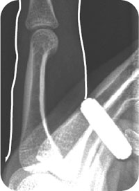
A radiograph shows changes of osteoarthritis at the DIP joint with full extension. Finger radiographs to obtain include posteroanterior, lateral (Fig. 47.1), and oblique views. A mallet finger results from injury to the extensor mechanism. On the lateral radiograph, the flexion deformity caused by lack of integrity of the extensor mechanism is clearly evident. The examiner should check this film for a flexion deformity at the DIP joint, with the distal phalanx flexed like a mallet. A tendinous or soft tissue mallet is an avulsion or tear of the distal extensor tendon at the DIP joint (Fig. 47.2). A bony mallet has an associated fracture of the dorsal base of the distal phalanx involving the insertion of the extensor tendon. A pure tendon injury shows no evidence of fracture, only the mallet deformity (Fig. 47.3).
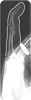
FIGURE 47.1 Lateral radiograph of the left hand of the patient in the introductory case, demonstrating soft tissue swelling over the left, second distal interphalangeal joint with a flexion deformity at that joint consistent with a mallet finger.
Related posts:


Stay updated, free articles. Join our Telegram channel

Full access? Get Clinical Tree


