Dushyant Purohit, MD, FRCPath, David M. Carpenter, PhD, and Thomas P. Naidich, MD
LEARNING OBJECTIVES
1. Appreciate the gyral and sulcal anatomy of the brain surface.
2. Appreciate the gross structure of the basal ganglia and thalami.
3. Appreciate the major commissural, projection, and association fiber tracts of the brain.
4. Appreciate the gross anatomy of the brainstem and cerebellum.
5. Appreciate the gross anatomy of the ventricular system.
6. Be able to identify those features in CT and MRI scans, in vivo.
This chapter presents first the anatomy of the meninges, the surface of the cerebral cortex, the deep gray nuclei, and the white matter tracts. It then illustrates how computed tomography (CT) and magnetic resonance (MR) imaging (I) display these structures. The chapter then addresses the anatomy and imaging of the brainstem, cerebellum, ventricles, and cisterns.
Gross Organization of the Brain
Adult brain weight varies from 1,000 to 1,700 grams (g) (fixed wet weight) and is typically larger in adult males (average 1,400 g) than adult females (average 1,275 g). From age 20 to 85, the average brain weight decreases by about 120 g. Approximately 90% of brain weight is cerebral, and 10% is cerebellar.
The skull base displays three major bone fossae (Fig. 2.1). From anterior to posterior, the anterior cranial fossa extends from the inner surface of the frontal bones to the posterior edges of the lesser wings of the sphenoid bones. It includes the orbital roofs, the cribriform plate, the planum sphenoidale, and the lesser wings of the sphenoid bones. The paired middle cranial fossae extend from the lesser and greater wings of the sphenoid bones anteriorly to the superior ridges of the petrous temporal bones posteriorly. On each side, the middle cranial fossae lie lateral to the sella turcica and the sloping upper surface of the tentorium. The posterior fossa lies posterior to the petrous temporal bones and clivus, inferior to the tentorium, and anterosuperior to the occipital bone. The posterior fossa communicates with the anterior and middle fossae through the arch-shaped opening in the tentorium designated the tentorial incisura (synonym: tentorial notch). The posterior fossa communicates with the spinal canal through the foramen magnum. Anatomically, portions of the brain and associated lesions are classified as supratentorial when they lie above the tentorium in the anterior and middle cranial fossae, as infratentorial when they lie below the tentorium within the posterior fossa, and as incisural when they occupy the tentorial notch.
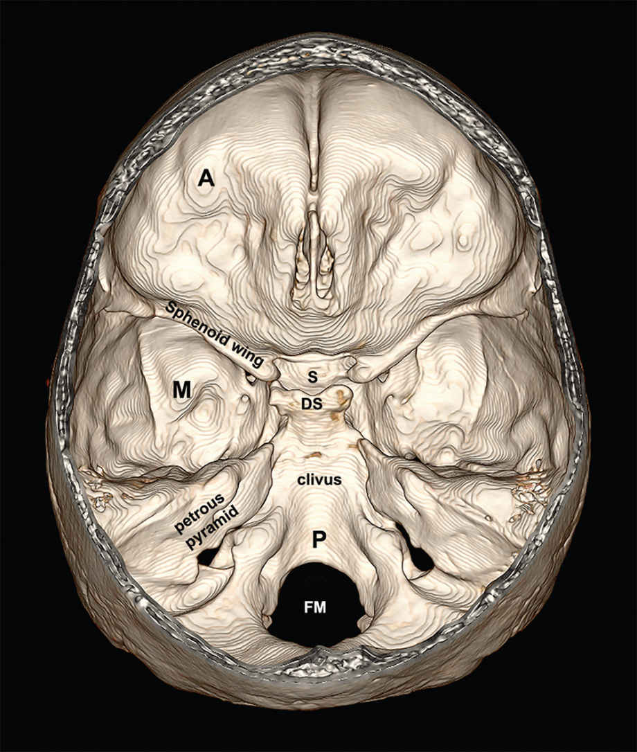
FIG. 2.1 Inner aspect of the skull base. Three-dimensional reconstruction from CT dataset with the calvarium removed. On each side, the sphenoid wing separates the anterior cranial fossa (A) from the middle cranial fossa (M). The superior ridge of the petrous pyramid separates the middle cranial fossa from the posterior fossa (P). The sella turcica (S) and dorsum sellae (DS) lie centrally. FM, foramen magnum.
Meninges
The brain proper is enclosed within three sets of meninges (Fig. 2.2A) (1–3). The outer, thick pachymeninges are termed dura mater. The dura has two layers, an outer periosteal layer and an inner meningeal layer. The outer periosteal layer adheres to the inner surface of each bone and turns outward into the sutures at the edges of each bone plate. The inner meningeal layer of the dura remains with the periosteal layer over most of the dural surface. In the midline and laterally, however, the inner meningeal layer turns sharply inward to form the falx and tentorium (Fig. 2.2B,C). The falx forms a sickle-shaped, midline fibrous partition that is shallow anteriorly and progressively deeper posteriorly. It attaches peripherally to the inner table of the skull in the midline. It attaches deeply to the crista galli anteriorly and the upper surface of the tentorium posteriorly, leaving a deep free margin between them. The left and right sides of the brain connect with each other through the falcine hiatus, inferior to the deep free margin of the falx.
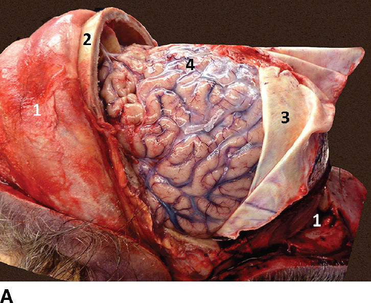
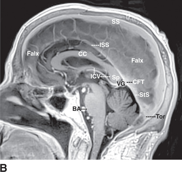
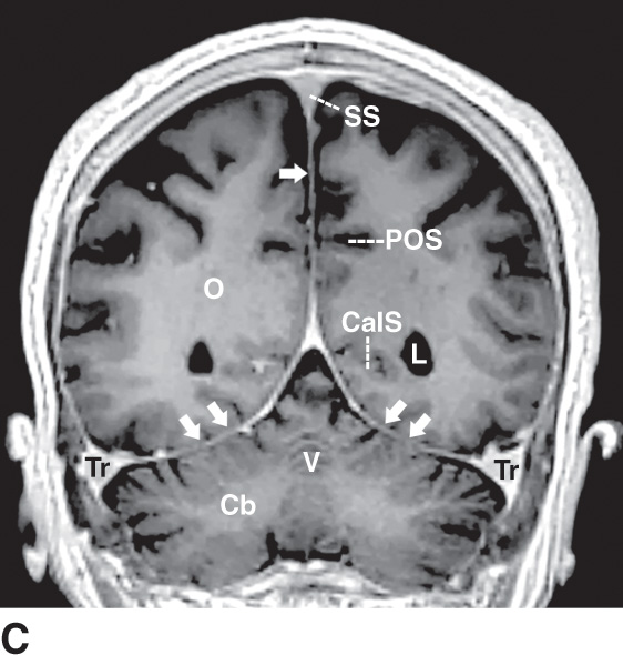
FIG. 2.2 Meninges. A: Lateral view. Fresh postmortem photograph after bicoronal skin incision; reflection of the skin, the galea aponeurotica, and the periosteum (1) both forward and back; and removal of much of the calvarium (2). The dura mater (3) has been reflected posteriorly to display the glistening arachnoid mater (4) overlying the brain. B,C: Dural partitions: falx and tentorium. B: Sagittal contrast-enhanced T1 image shows the sickle shape of the midline falx, the superior sagittal sinus (SS) at the periphery of the falx, the inferior sagittal sinus (ISS) along the deep free margin of the falx, and the straight sinus (StS) along the confluence of the posterior falx and tentorium (CFT). These join together at the confluence of the sinuses (torcular herophili) (Tor). The paired internal cerebral veins (ICV) drain through the vein of Galen into the straight sinus. BA, basilar artery; CC, corpus callosum; Sp, splenium of the corpus callosum. C: Coronal contrast-enhanced T1 image shows the tent-like configuration of the tentorium (paired dual arrows) and the midline falx (single arrow). The cerebrum lies in the supratentorial compartment above the tentorium, and the cerebellum (Cb) lies within the infratentorial compartment below. The superior sagittal sinus (SS), straight sinus, and transverse sinuses (Tr) are enclosed within the walls of the falx and tentorium. The calcarine sulcus (CalS) is the first long, horizontally oriented sulcus above the tentorium and is directed at the medial edge of the lateral ventricles (L). The parieto-occipital sulcus (POS) is also horizontally oriented and lies above the CalS. O, occipital lobe; V, vermis.
The tentorium is tent shaped. Posteriorly and laterally, the left and right sides of the tentorium attach to the occipital bone, swing forward and medially along the superior petrous ridges, and continue on to the posterior clinoid processes of the sella as the petroclinoid ligaments. Medially, the tentorium attaches to the undersurface of the posterior falx and then forms paired free medial margins that sweep forward to reach the anterior clinoid process on each side. The supratentorial and infratentorial portions of the brain connect with each other through the incisura between the free medial margins of the tentorium. On imaging studies, the falx and tentorium appear as sharply defined contrast-enhancing dural reflections (Fig. 2.2B,C).
Veins enclosed at the deep and superficial margins of the two layers of meningeal dura become the dural venous sinuses (Fig. 2.2B,C) (1–3). The superior sagittal sinus lies at the peripheral margin of the falx. The inferior sagittal sinus lies along the free deep margin of the falx. The straight sinus runs in the midline along the confluence of the posterior falx and tentorium. The paired transverse sinuses course laterally along the posterolateral margins of the tentorium, turn down behind the lateral edges of the petrous pyramids as the paired sigmoid sinuses, and merge into the paired jugular veins. The superior sagittal sinus, straight sinus, and paired transverse sinuses converge in the midline posteriorly to form the variably complete confluence of the sinuses (torcular herophili). Blood normally flows from anterior to posterior in the superior sagittal sinus, inferior sagittal sinus, and straight sinus; converges in the midline posteriorly at the torcular; and then flows laterally, to each side, along the paired transverse sinuses to reach the jugular veins. Deep to the dura, the thin leptomeninges comprise the arachnoid mater and pia mater. Cerebrospinal fluid (CSF) fills the subarachnoid space deep to the arachnoid mater and external to the pia mater. The pia and arachnoid typically show no appreciable contrast enhancement.
Supratentorial Brain
Surface anatomy
The cerebrum is organized into two hemispheres. Each hemisphere has a rounded lateral surface (the convexity) that faces the inner surface of the calvarium, a flat medial surface that faces the falx and the opposite hemisphere, and a generally concave inferior surface that faces the skull base and tentorium (Figs. 2.3 and 2.4). These surfaces meet superiorly at the superior margin of the brain, inferolaterally at the inferior margin, inferomedially at the tentorial margin, and anteriorly at the frontal (orbital) margin. The pointed extremities of each hemisphere are designated poles: hence frontal poles anteriorly, temporal poles anteroinferiorly, and occipital poles posteriorly.
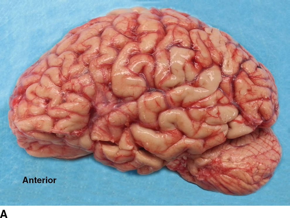
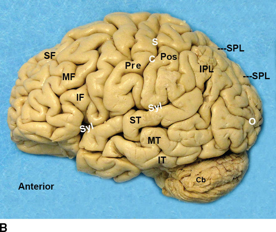
FIG. 2.3 Convexity surface of the left hemisphere. Lateral views. A: The fresh (unfixed) specimen shows the pia–arachnoid and vessels overlying the gyri and sulci of the brain. B: The same specimen after formalin fixation and removal of the meninges and vessels. The central sulcus (CS) separates the superior frontal (SF), middle frontal (MF), inferior frontal (IF), and precentral (Pre) gyri of the frontal lobe from the postcentral gyri (Pos), inferior parietal lobule (IPL), and superior parietal lobule (SPL) of the parietal lobe. The sylvian fissure (Syl) separates the frontal and parietal lobes above from the superior temporal (ST), middle temporal (MT), and inferior temporal (IT) gyri of the temporal lobe below. On the convexity, the occipital lobe (O) is poorly demarcated from the temporal and parietal lobes. The cerebellum (Cb) lies below.
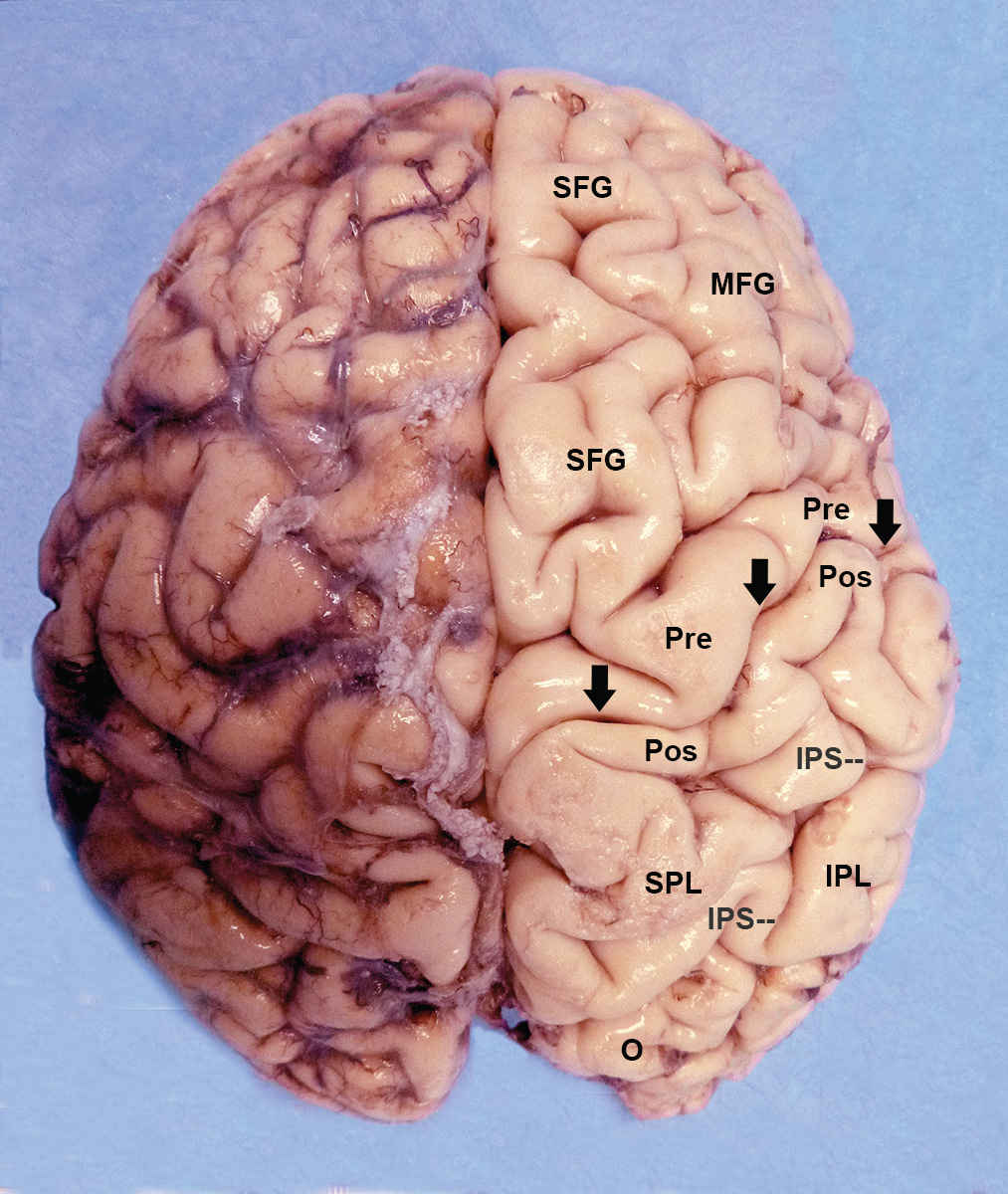
FIG. 2.4 Superior surface of the fixed brain. Unilateral removal of the meninges and vessels exposes the gyri and sulci beneath. The central sulcus (black arrows) separates the superior (SFG), middle (MFG), and precentral (Pre) gyri of the frontal lobe from the postcentral gyrus (Pos), the superior parietal lobule (SPL), and inferior parietal lobule (IPL) of the parietal lobe. The intraparietal sulcus (IPS) divides the superior parietal lobule superomedially from the inferior parietal lobule inferolaterally. O, occipital lobe.
Anatomically, the cerebrum is divided into lobes, lobules, and gyri (1–3). Classically, there are 5 lobes on each side: the paired frontal, parietal, temporal, occipital, and insular lobes. A sixth, single “limbic lobe” is conceptualized as paired arcs of tissue from both hemispheres that, together, form a ring (limbus) around the brainstem at the central medial edge of the brain. The frontal, parietal, temporal, and occipital lobes are visible on both the lateral (convexity) surface and the medial surface. The insular lobe is buried within the sylvian fissure, deep to the convexity (Fig. 2.5). The limbic lobe is seen only on the inferomedial surface of the brain.
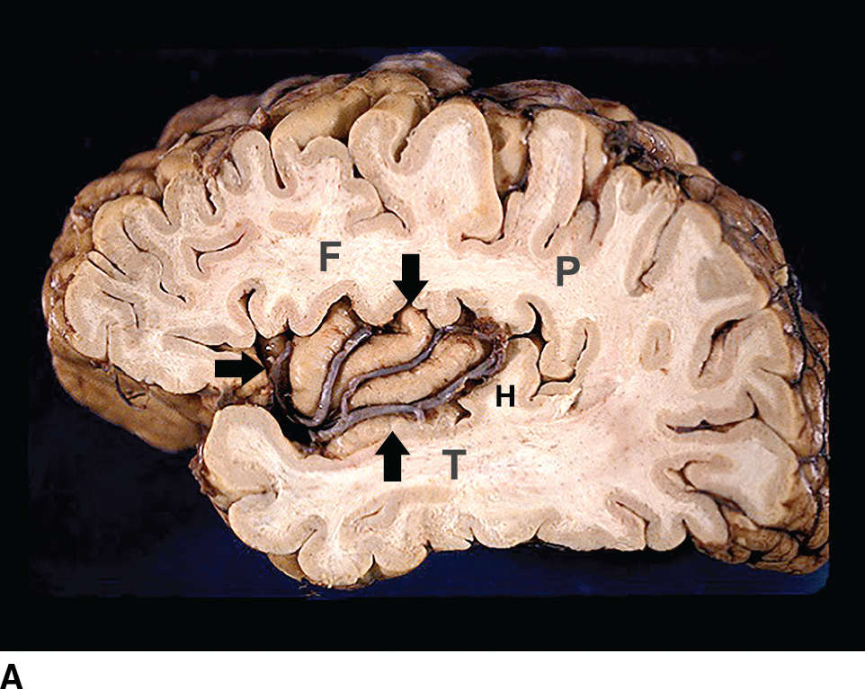
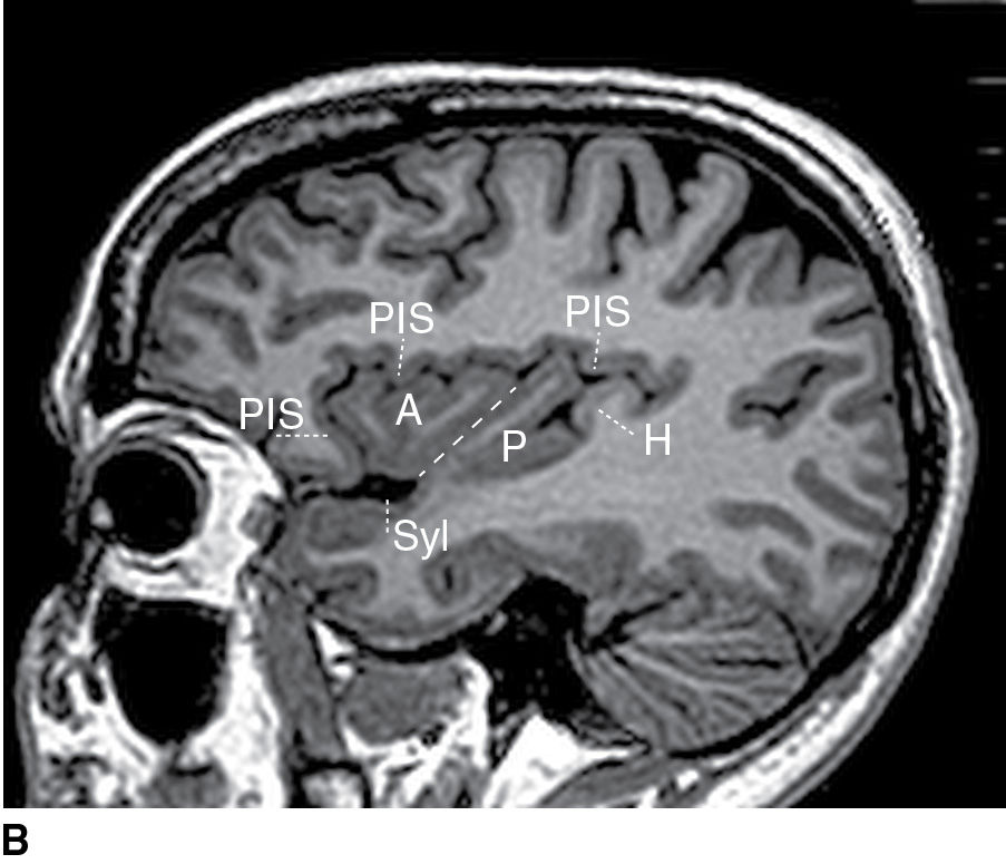
FIG. 2.5 Insula. A: Fixed postmortem specimen. Lateral view. Resection of the convexity portions of the frontal (F), parietal (P), temporal (T), and occipital lobes reveals the triangular peri-insular sulcus (arrows) and the M2 segments of the middle cerebral artery coursing over the lateral surface of the insula. H, Heschl transverse temporal gyrus. B: Sagittal noncontrast T1 MRI displays the sylvian fissure (Syl), the peri-insular sulcus (PIS), the central sulcus (dashed line) separating the anterior (A) and posterior (P) lobules of the insula, and Heschl gyrus (H).
Convexity (lateral) surface
On the convexity, the major structures are demarcated by the natural landmarks of the sylvian fissure, the central sulcus, and the intraparietal sulcus (1–4). The posterior temporal lobe and posterior parietal lobe are poorly demarcated from the anterior occipital lobe. Anatomically and functionally, therefore, it is often more useful to consider the junction of the temporal (T), parietal (P), and occipital (O) lobes holistically as a “TPO confluence.”
Frontal lobe
The convexity surface of the frontal lobe extends back from the frontal pole to the central sulcus, above the sylvian fissure (Figs. 2.3 and 2.4). From superior to inferior, the frontal convexity displays three “horizontal” gyri: the long superior frontal gyrus (SFG), the large undulating middle frontal gyrus (MFG), and the small triangular inferior frontal gyrus (IFG). These are separated by the superior and inferior frontal sulci. The large MFG is often subdivided into superior and inferior halves by a horizontal middle frontal sulcus. Just posterior to the three horizontal gyri, the frontal convexity displays an additional vertical precentral gyrus (preCG) situated behind the precentral sulcus and immediately anterior to the central sulcus. The preCG contains the primary motor cortex (designated M1). The cortex of the preCG is notably thicker than is the cortex of the postcentral gyrus.
Parietal lobe
The convexity surface of the parietal lobe extends back from the central sulcus to the poorly demarcated TPO confluence (Figs. 2.3 and 2.4). Anteriorly, the parietal lobe has one vertical postcentral gyrus situated immediately behind the central sulcus and anterior to the postcentral sulcus. The postcentral gyrus contains the primary somatosensory cortex (designated S1). The cortex of the postcentral gyrus is notably thinner than the cortex of the preCG. Posteriorly, the parietal lobe is subdivided into a superior parietal lobule and an inferior parietal lobule by a deep, arcuate, intraparietal sulcus.
Temporal lobe
The convexity surface of the temporal lobe lies inferior to the sylvian fissure and extends back from the temporal pole to the TPO confluence. Like the frontal lobe, the temporal convexity displays three horizontal gyri: the superior temporal gyrus, middle temporal gyrus, and inferior temporal gyrus, separated by the superior and inferior temporal sulci (Fig. 2.3). The inferior temporal gyrus forms the inferior margin of the temporal convexity and, without changing name, curves under the margin to form the most lateral gyrus of the inferior temporal surface.
Occipital lobe
The convexity surface of the occipital lobe extends backward from the parietal and temporal lobes to the occipital pole (Fig. 2.3). The convexity surface of the occipital lobe is small, curves far medially to reach the midline, and appears markedly foreshortened in true lateral images. It displays three horizontal gyri: the superior, the middle, and the inferior occipital gyri, separated by the superior and inferior occipital sulci. The large middle occipital gyrus may be subdivided into superior and inferior halves by a middle occipital sulcus.
Insula (insular lobe)
The insulae are vertically oriented, laterally convex, shell-like lobes that lie deep within the sylvian fissures beneath the overhanging frontoparietal and temporal opercula (lips) (2,5). On each side, the inferior end of the central sulcus runs obliquely across the insula, dividing the insula into anterior and posterior insular lobules (Fig. 2.5). The insular segments of the middle cerebral arteries (designated M2 segments) course over the surface of the insula.
Medial and inferomedial surfaces
The medial surface of the brain displays a radial organization, with the gyri and sulci organized around the corpus callosum as a series of concentric, cocurvilinear arcs and a set of radians that are oriented “perpendicular” to these arcs (Figs. 2.6 and 2.7; Table 2.1) (1–3).
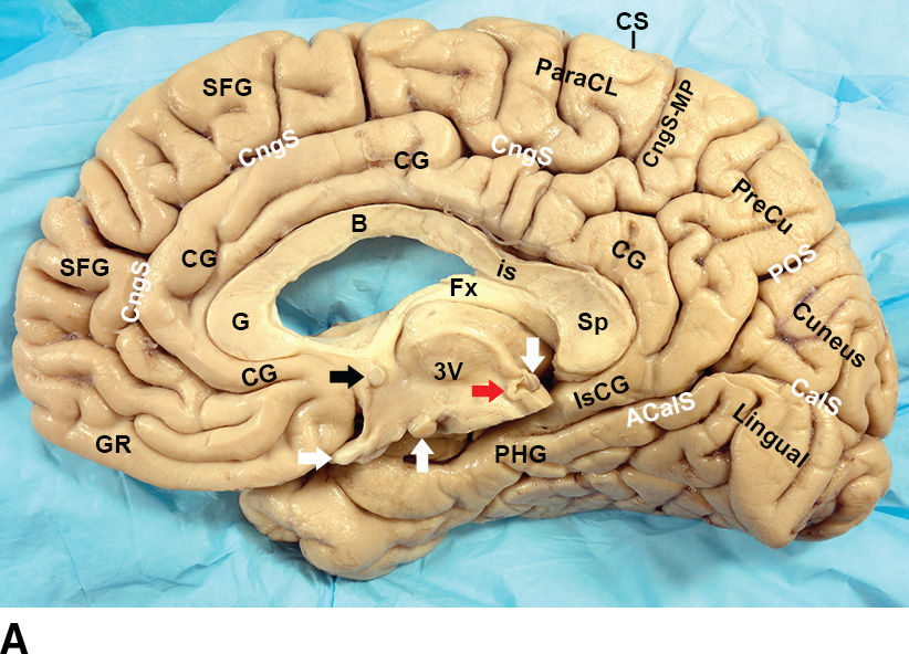
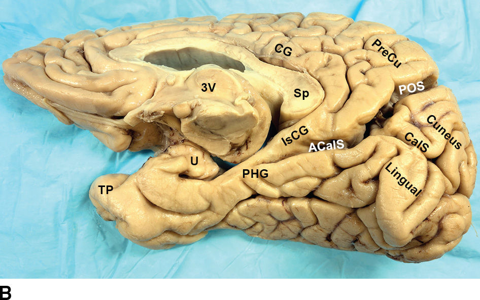
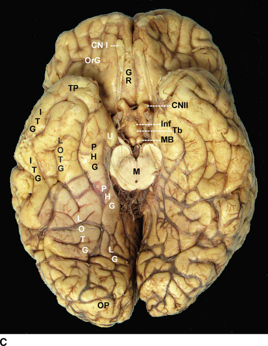
FIG. 2.6 Medial and inferior surfaces of cerebrum. A,B: Medial and inferomedial views of the fixed brain. A: Centrally, the specimen displays the third ventricle (3V), the optic chiasm (horizontal white arrow), mammillary body (up-pointing white arrow), anterior commissure (black arrow), posterior commissure (red arrow), pineal gland (down-pointing white arrow), and fornix (Fx). In progressively more peripheral layers, it displays the genu (G), body (B), isthmus (Is), and splenium (SP) of the corpus callosum, the cingulate gyrus (CG), the cingulate sulcus (CngS), the marginal segment of the cingulate sulcus (CngS-MP, pars marginalis), the gyrus rectus (GR), the medial surface of the superior frontal gyrus (SFG) and the paracentral lobule (ParaCL). Posteriorly, it shows the “lazy Y” formed by the parieto-occipital sulcus (POS), the calcarine sulcus (CalS), and the anterior calcarine sulcus (ACalS). The parahippocampal gyrus (PHG) forms the medial edge of the temporal lobe. B: Rotating the brain to expose more of the inferior surface shows the relations of the isthmus of the cingulate gyrus (IsCG) to the splenium (Sp) and the anterior calcarine sulcus (ACalS) and how the parahippocampal gyrus (PHG) passes forward and then hooks backward, superiorly, and medially to form the uncus (U). TP, temporal pole. C: Base view of a second fixed specimen demonstrating the orbital surface of the frontal lobes and the inferior surface of the temporo-occipital lobes. (See Table 2.1 for definitions of labels.)
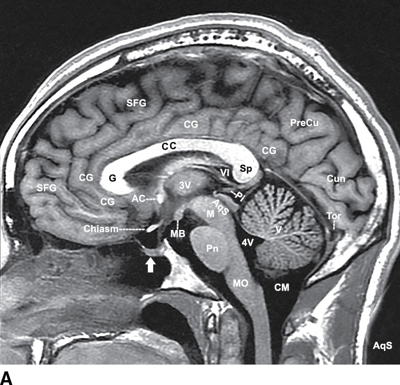
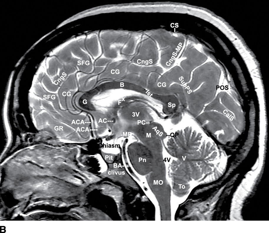
FIG. 2.7 Midsagittal T1 (A) and T2 (B) MR images. White arrow in (A) points to the sella turcica/pituitary gland. (See Table 2.1 for definitions of labels.)
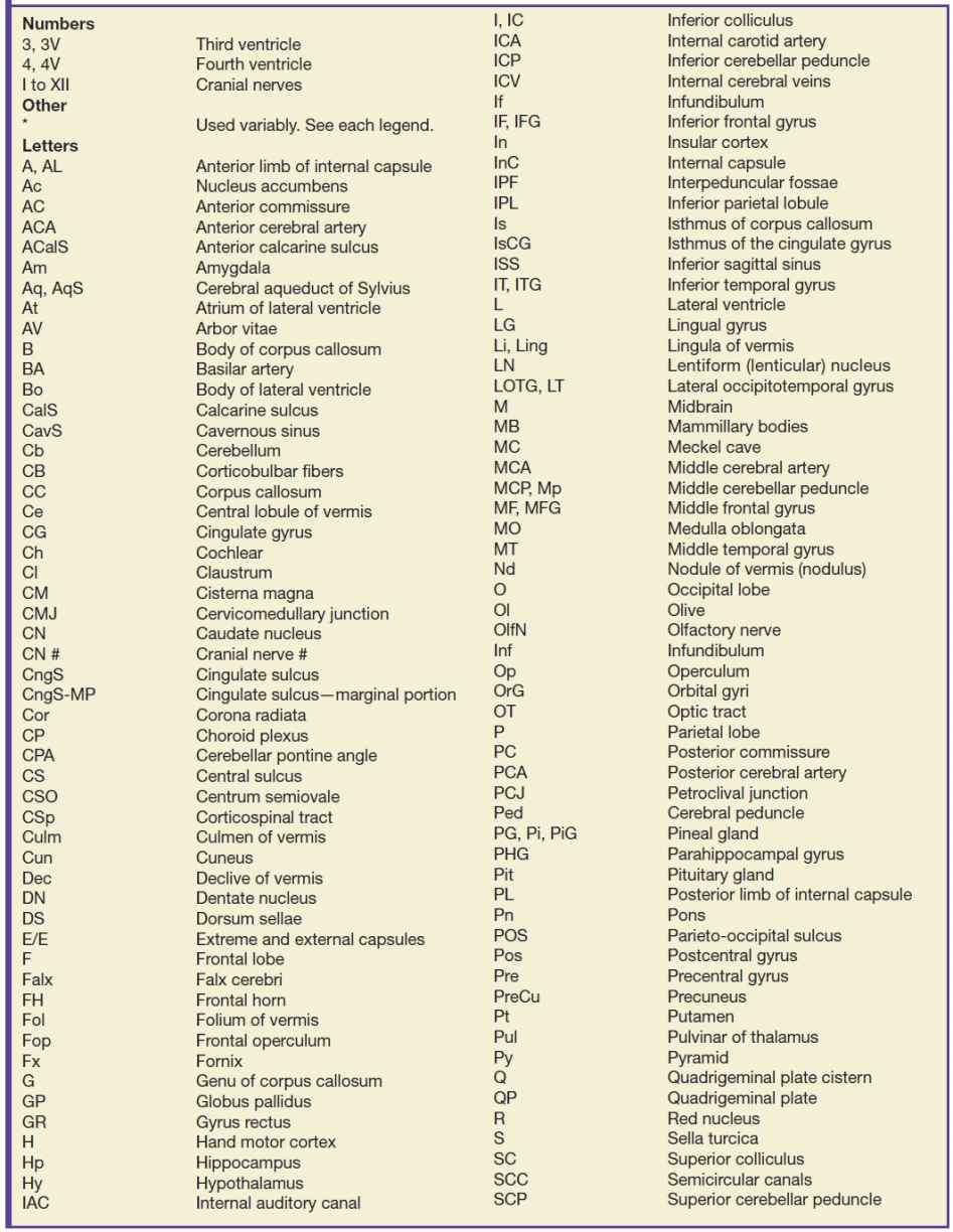
Table 2.1 ABBREVIATIONS USED IN FIGURES
Anteriorly, from central to superficial, the key natural landmarks of the medial surface include the corpus callosum, callosal sulcus, cingulate gyrus, cingulate sulcus, and the marginal portion of the cingulate sulcus (pars marginalis) (Figs. 2.6 and 2.7). The uppermost end of the central sulcus typically notches the superior margin of the cerebral hemisphere shortly in front of the pars marginalis. The tissue surrounding the uppermost end of the central sulcus on the medial surface is the paracentral lobule. The paracentral lobule is defined by the paracentral sulcus anteriorly and the pars marginalis posteriorly. Posterosuperiorly, the key landmarks include the subparietal sulcus, the parieto-occipital sulcus, the calcarine sulcus, and the anterior calcarine sulcus (Figs. 2.6 and 2.7).
Frontal lobe
The medial surface of each frontal lobe lies peripheral to the cingulate sulcus. It includes the gyrus rectus, the medial aspect of the SFG (sometimes called the medial frontal gyrus), and the anterior portion of the paracentral lobule (Figs. 2.6 and 2.7). The inferior surface of the frontal lobe overlies the orbit. It displays the gyrus rectus medially, the long olfactory sulcus just lateral to the gyrus rectus, and a group of orbital gyri situated lateral to the olfactory sulcus. The olfactory bulb and tract overlie the olfactory sulcus.
Parietal lobe
The medial surface of the parietal lobe is defined by the central sulcus anteriorly, the parieto-occipital sulcus posteriorly, and the subparietal sulcus inferiorly (Figs. 2.6 and 2.7). It includes two gyri: (i) the posterior portion of the paracentral lobule situated between the paracentral sulcus and the pars marginalis and (ii) the precuneus situated behind the pars marginalis, anterior to the parieto-occipital sulcus and superior to the subparietal sulcus.
Occipital lobe
The medial surface of the occipital lobe will be considered in two sections. (i) The posterior superior portion of the occipital lobe is defined by the union of the parieto-occipital sulcus with the calcarine sulcus to form the anterior calcarine sulcus. This union resembles a capital letter Y tilted on its side (the “lazy Y” sign) (Figs. 2.6 and 2.7). The precuneus lies anterior to the parieto-occipital sulcus. The cuneus lies behind the parieto-occipital sulcus above the calcarine sulcus. The lingual gyrus (synonym: medial occipitotemporal gyrus) lies inferior to the calcarine sulcus behind the anterior calcarine sulcus. The cingulate gyrus curves behind the splenium and narrows to the isthmus of the cingulate gyrus anterior to the anterior calcarine sulcus. (ii) The more inferior occipital lobe merges with the temporal lobe, discussed next.
Temporal and occipital lobes
Because the temporal and occipital lobes merge together and slope down over the tentorium, they display a confluent inferomedial surface (Figs. 2.6 and 2.7) (1–3). Anteriorly, from lateral to medial, the inferomedial surface of each temporal lobe includes the inferior temporal gyrus, the lateral occipitotemporal gyrus, and the parahippocampal gyrus, separated by the lateral occipitotemporal sulcus and the collateral sulcus. Three temporal gyri deserve special attention. Medially, the parahippocampal gyrus forms the medial-most edge of the temporal lobe along the full length of the temporal lobe. The anterior end of the parahippocampal gyrus hooks sharply posteriorly, superiorly, and medially to form the uncus (Fig. 2.6B). Superiorly, the transverse temporal gyrus of Heschl (Heschl gyrus) courses obliquely across the upper surface of the temporal lobe. Heschl gyrus is the primary auditory cortex (designated A1). Inferiorly, the lateral occipitotemporal gyrus runs the full length of the brain from the temporal lobe into the occipital lobe. The term fusiform gyrus is used variably but typically indicates a central portion of the lateral occipitotemporal gyrus spanning both lobes. It is considered a “face recognition” region.
Hippocampal formation
Medially, the temporal lobe displays the hippocampal formation (Fig. 2.8) (1–3,6). The hippocampal formation is the folded medial temporal cortex that lies superomedial to the parahippocampal gyrus along the full length of the medial wall of the temporal horn. The hippocampal formation consists of both gray matter and white matter. The gray matter components are the subiculum inferiorly, the dentate gyrus superiorly, and the hippocampus per se (Ammon horn) superolaterally. The white matter components are the alveus and fimbria, which together form the fornix. The alveus is the layer of white matter situated between the ependyma of the temporal horn and the hippocampus per se. This layer of white matter curves medially to form a free edge of white matter designated the fimbria. The alveus and fimbria pass posteriorly, gather progressively more fibers, and thicken progressively to constitute the fornix.
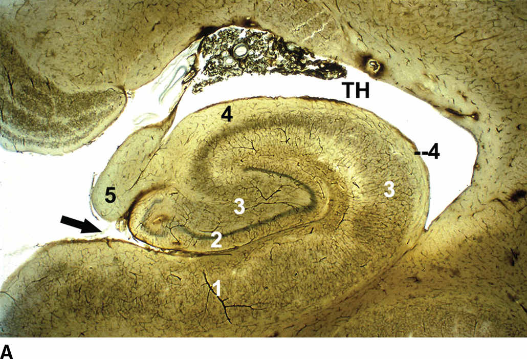
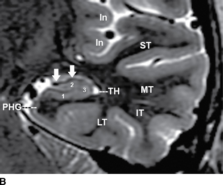
FIG. 2.8 The hippocampal formation. A: Coned-down histologic section of the hippocampal formation. B: Coned-down coronal T2 MR image of the anterior temporal lobe. The hippocampal formation forms the inferomedial wall of the temporal horn (TH). It includes the subiculum (1), dentate gyrus (2), and hippocampus (3) arrayed above and below the hippocampal fissure (black arrow in A). The white matter fibers of the hippocampal formation pass to the alveus (4) and fimbria (5) (white arrows in B), forming the fornix. The surface of the anterior temporal lobe shows 5 gyri: superior temporal gyrus (ST), middle temporal gyrus (MT), inferior temporal gyrus (IT), lateral occipitotemporal gyrus (LT), and parahippocampal gyrus (PHG). The parahippocampal gyrus forms the medial-most surface of the temporal lobe, immediately inferomedial to the hippocampal formation. The inferior temporal gyrus forms the inferolateral margin of the temporal lobe. In, insula. (A, courtesy of Dr. Dixon Moody.)
Limbic lobe
The limbic lobe includes the cingulate gyri situated between the callosal and cingulate sulci, the isthmi of the cingulate gyri situated beneath the splenium and superior to the anterior calcarine sulci, and the parahippocampal gyri situated along the full medial margins of the temporal lobes. The inferior portion of the limbic lobe, encircling the midbrain, is shown in Figure 2.6C.
Deep gray matter
The term deep gray matter is used to designate all of the supratentorial gray matter deep to the cerebral cortex, plus selected infratentorial structures. It includes the multiple subdivisions of the basal ganglia and the thalami (Figs. 2.9 to 2.13) (1–3).
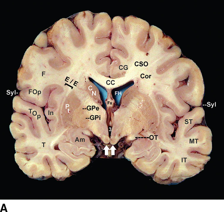
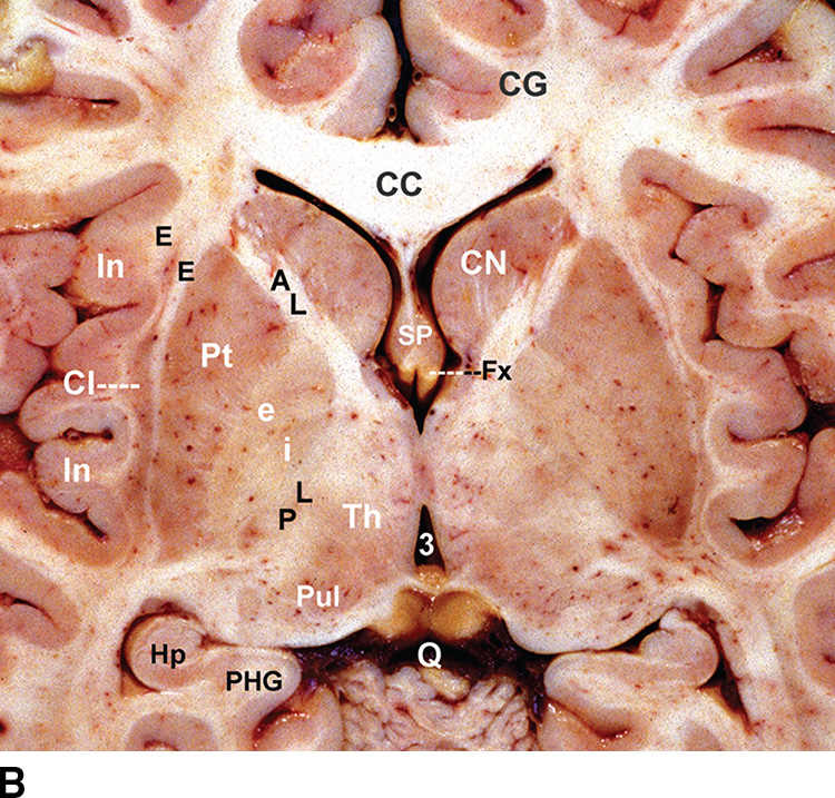
FIG. 2.9 Deep gray matter. Coronal (A) and magnified axial (B) anatomic sections through the frontal operculum (FOp), temporal operculum (TOp), sylvian fissure (Syl), insula (In), external and extreme capsules (E/E), putamen (Pt), globus pallidus (GP) externa (e) and interna (i), internal capsule (AL, anterior limb; PL, posterior limb), caudate nucleus (CN), thalamus (Th), and amygdala (Am). Centrally, one finds the paired frontal horns (FH), septum pellucidum (SP), anterior columns of the fornix (Fx), third ventricle (3), mammillary bodies (white arrows), and optic tracts (OT). (See Table 2.1 for definitions of labels.)
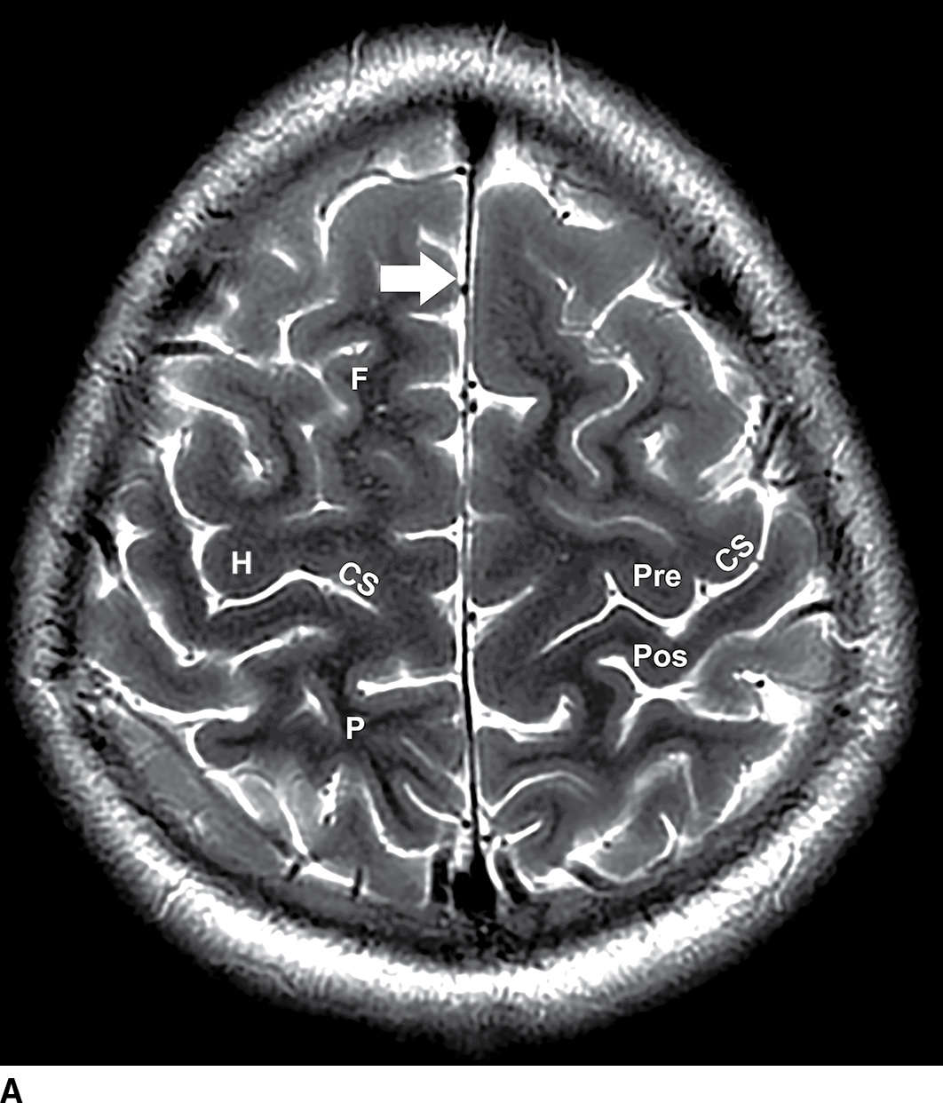
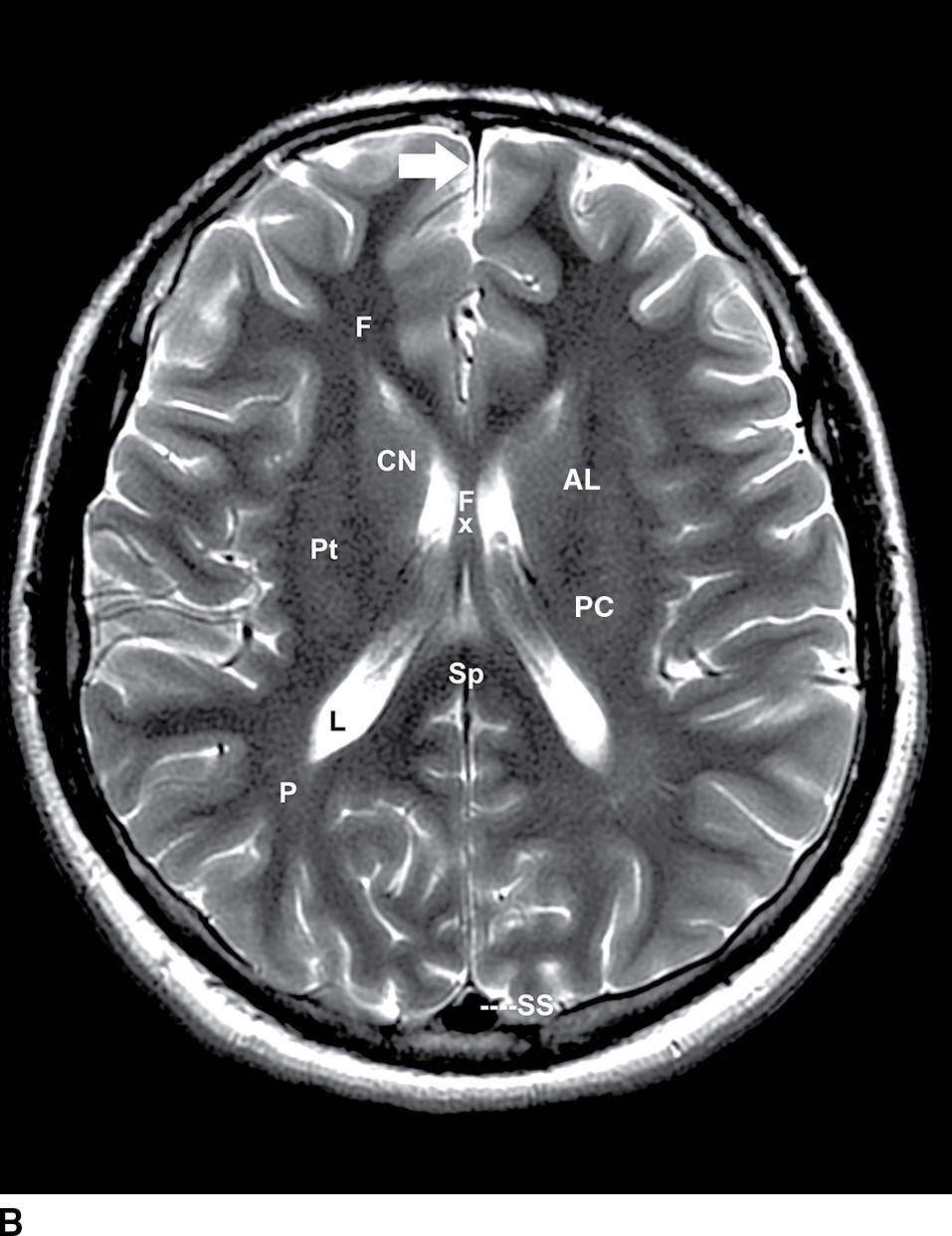
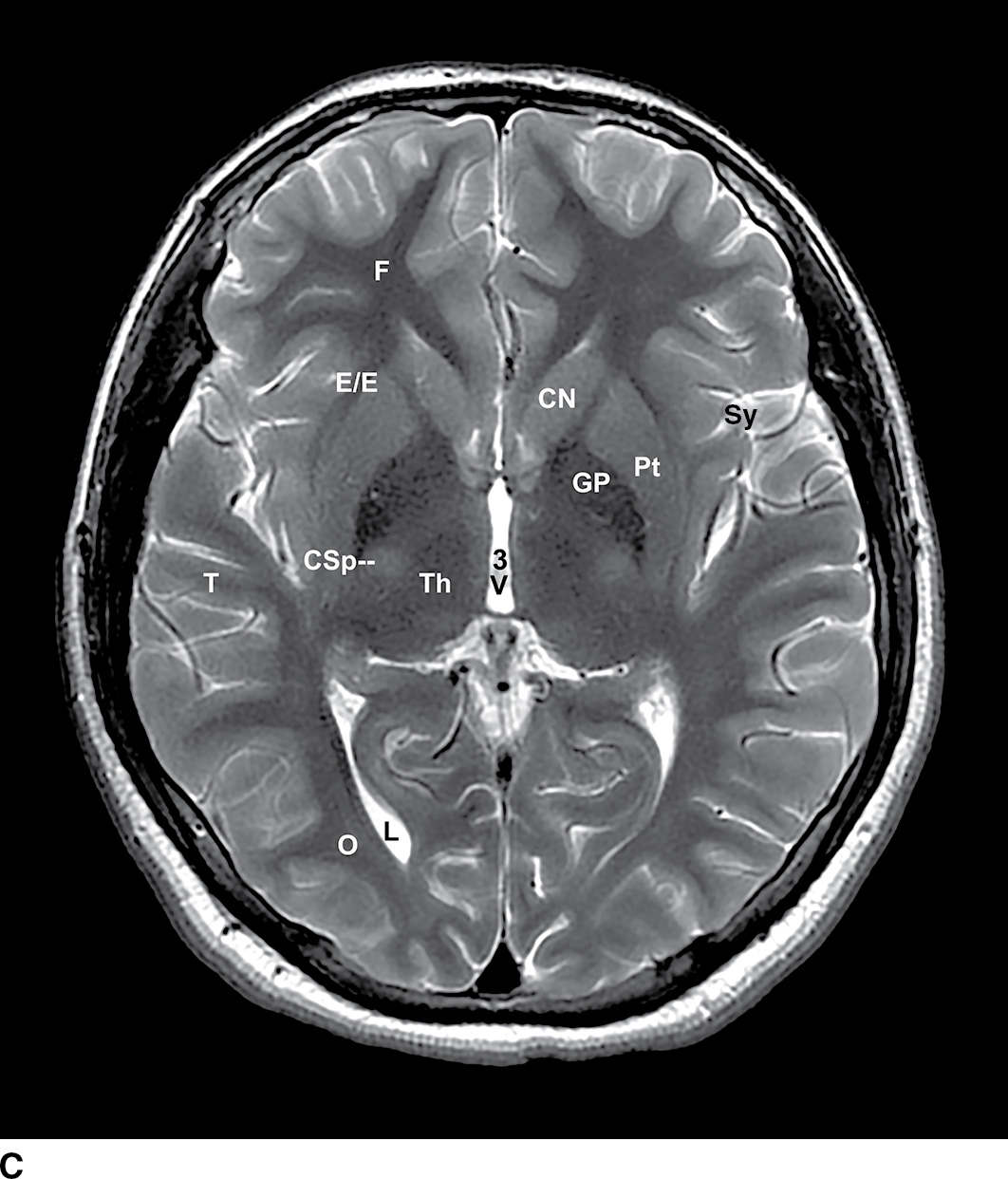
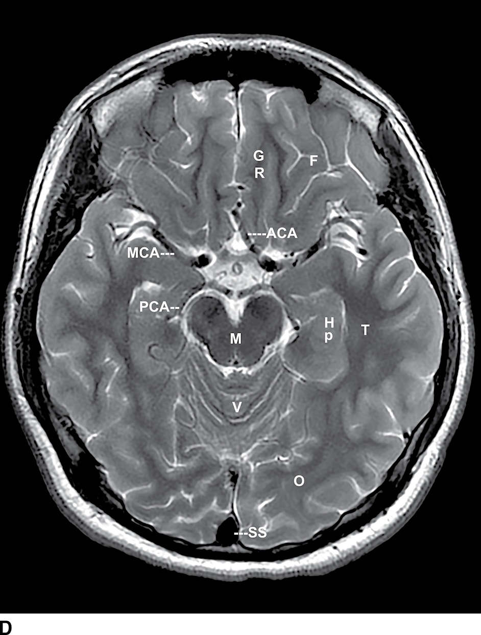
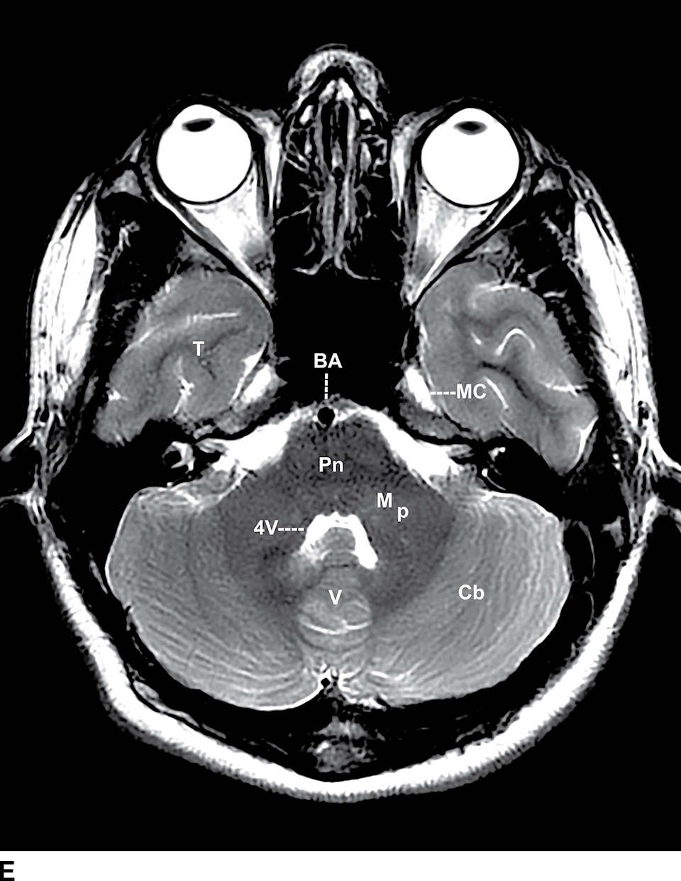
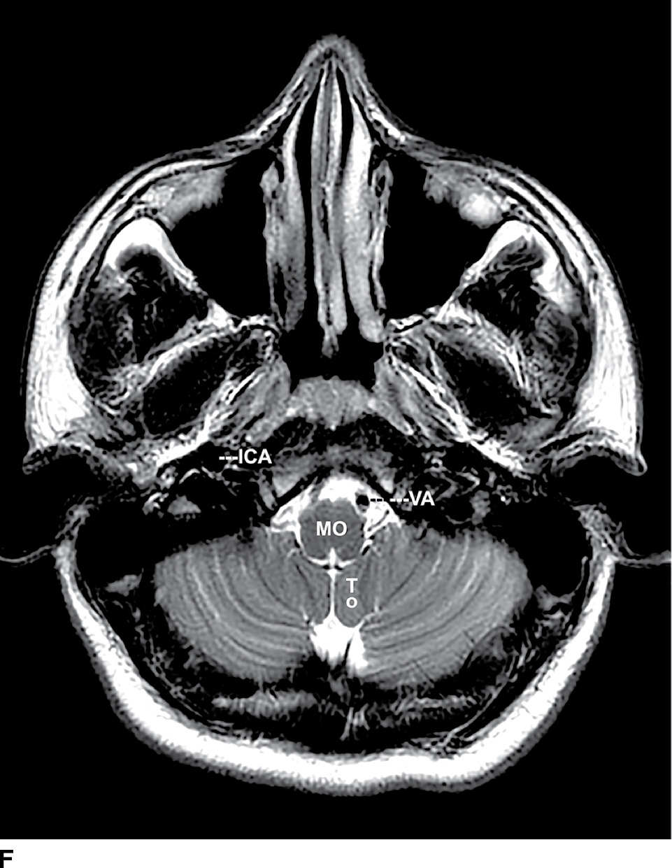
FIG. 2.10 Noncontrast axial T2 MRI of the brain. From superior to inferior, selected sections demonstrate the structures shown by T2 MRI at the level of the vertex (A), lateral ventricles (B), third ventricle (C), midbrain (D), pons (E), and medulla (F). Arrows indicate the falx and tentorium. (See Table 2.1 for definitions of labels.)
Related posts:


Stay updated, free articles. Join our Telegram channel

Full access? Get Clinical Tree


