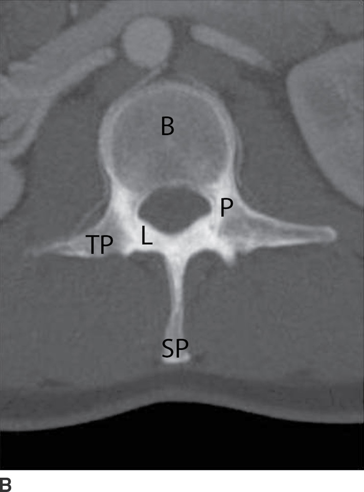
FIG. 10.1 A: Sagittal CT MIP of the lumbar spine demonstrating the vertebral body (B), pedicle (P), superior articular process (SAP), inferior articular process (IAP), and zygapophyseal joint (Z). The superior articular processes extend superiorly from the junction of the laminae and pedicles, while the inferior articular processes extend inferiorly from the undersurface of each lamina. The zygapophyseal joints are formed by the superior articular facet and the inferior articular facet. The pars interarticularis (PI) is the portion of the lamina between the superior and inferior articular processes. The intervertebral foramen (F) allows for the passage of spinal nerves and vessels. B: Axial CT MIP of the lumbar spine demonstrating the vertebral body (B) and posterior neural arch. (P, pedicle; TP, transverse process; L, lamina; SP, spinous process.)
The intervertebral disks consist of the inner nucleus pulposus and the outer annulus fibrosis. The main function of the disks is to distribute load and to allow flexion/extension and lateral bending. A remnant of the notochord, the nucleus pulposus, is usually located more dorsally compared to the center of the vertebral body. The annulus fibrosis attaches to the anterior longitudinal ligament (ALL) and posterior longitudinal ligament (PLL) and is also fused to the epiphyseal ring of the vertebral bodies. MRI of a normal disk demonstrates high signal on T2-weighted imaging related to the water content of the nucleus pulposus and inner annulus fibrosis. The outer annulus fibrosis demonstrates low signal on T1- and T2-weighted imaging (2) (Fig. 10.2).
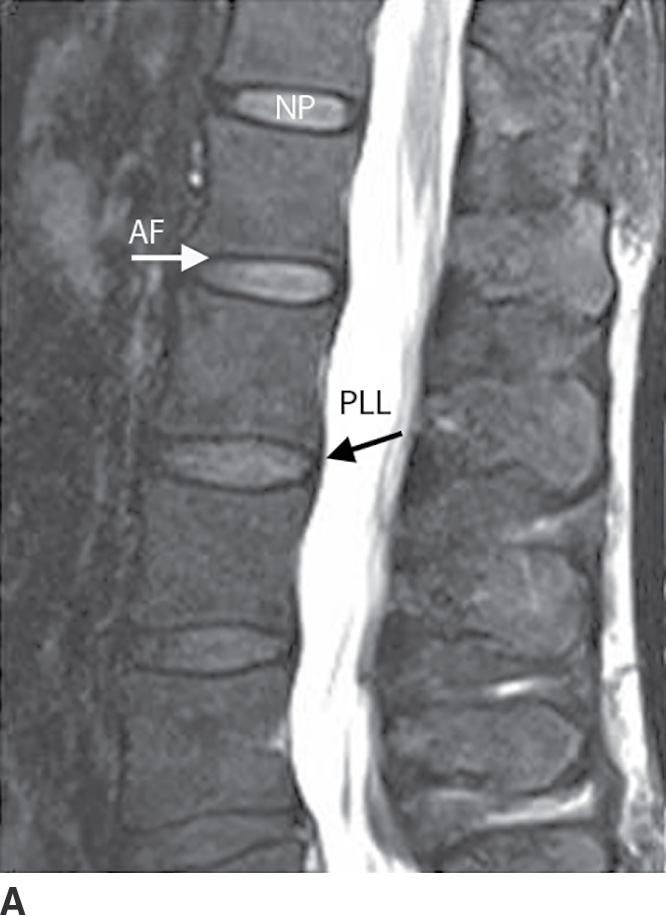
FIG. 10.2 Sagittal T2 fat-saturated (A) and axial T2-weighted (B) MRIs of the lumbar spine demonstrating high signal intensity in the nucleus pulposus (NP) and inner annulus fibrosis and decreased signal in the outer annulus fibrosis (AF). The anterior longitudinal ligament (ALL) is not well seen, but the posterior longitudinal ligament (PLL) is seen as a continuous band of signal hypointensity along the posterior vertebral body. The ligamentum flavum (LF) is also seen posteriorly in the spinal canal.
Cervical spine
There are seven cervical vertebrae. The first and second cervical vertebrae are unique. The first cervical vertebra (axis or C1) has no body or spinous process and consists of an anterior and posterior arch. On the ventral surface of the anterior arch of C1 is the anterior tubercle, which serves as the attachment for the longus coli muscles. The dorsal concave aspect of the anterior arch articulates with the odontoid process of C2. Joining the anterior and posterior arches are the rather large lateral masses. The cup-shaped superior articular processes articulate with the occipital condyles to support the weight of the head. The flattened inferior articular processes articulate with C2, allowing for head rotation (1) (Fig. 10.3).
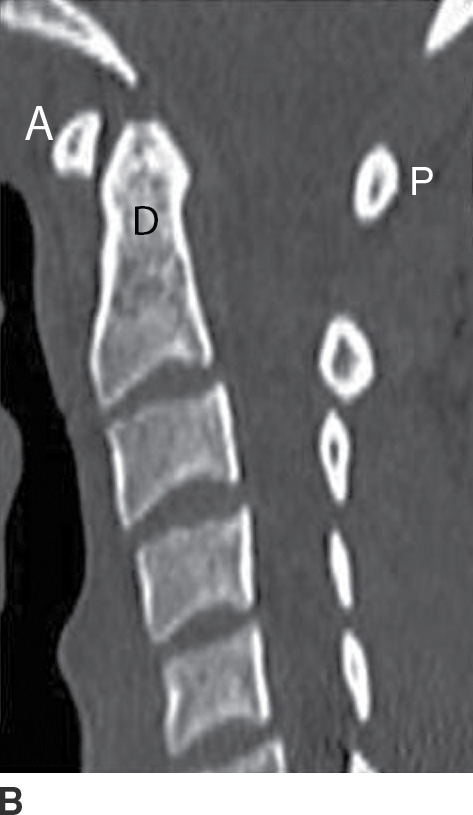
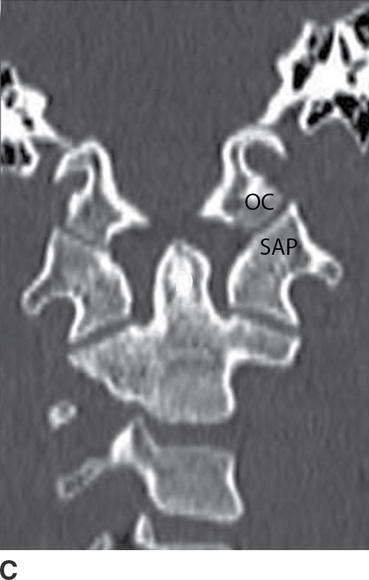
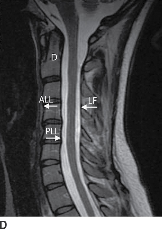
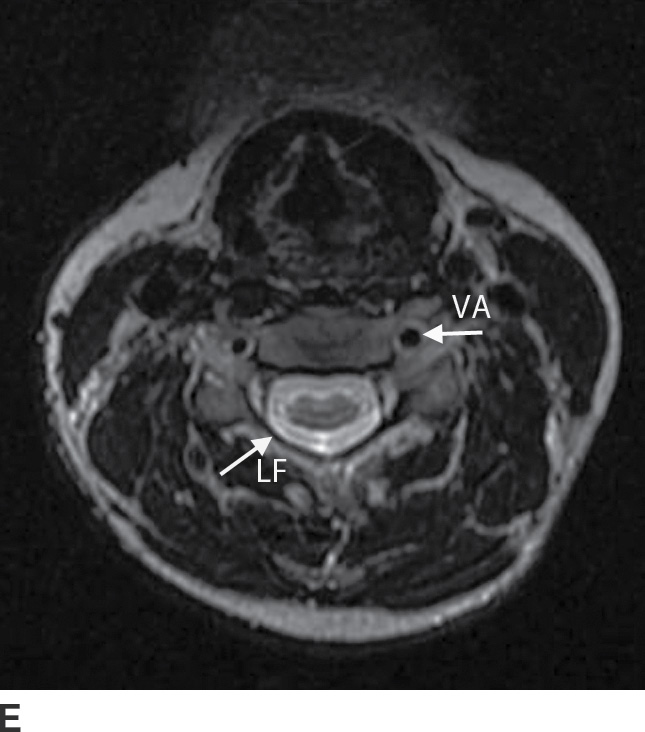
FIG. 10.3 Axial (A), sagittal (B), and coronal (C) cervical spine CTs demonstrating the anterior (A) and posterior (P) arches of C1 as well as the foramen transversarium (FT). Note the anterior tubercle (white arrow). Although the vertebral artery is not seen, its course runs through the arcuate foramen (AF), whose floor is present bilaterally. The cup-shaped superior articular processes (SAP) articulate with the occipital condyles (OC). The dens (D) is seen extending from the body of C2. Two small tubercles (black arrow) are seen on the medial borders of C1, serving as anchors for the transverse ligament, which secures the dens and allows for lateral rotation of C1 on C2. Sagittal (D) and axial (E) T2-weighted MRIs demonstrating the anterior (ALL) and posterior (PLL) longitudinal ligaments and the ligamentum flavum (LF). The vertebral artery (VA) flow voids are seen within the foramen transversarium.
The axis, or second cervical vertebra, forms from four ossification centers: two neural arches, the body, and the odontoid process. The dens, or odontoid process, projects superiorly from the body of the axis and serves as a pivot for the atlas (1). The os terminale is an additional ossification center at the tip of the dens that fuses with the odontoid process by age 3 to 6 (3). The pedicles and lamina are strong, and the vertebral canal is wide at this level, although smaller than at C1. The superior articular surfaces are rounded, directed superiorly and laterally, and supported by the body, pedicles, and transverse processes; the inferior articular surfaces assume a similar orientation to the adjacent lower vertebral body. The transverse processes end in a single tubercle and are small. The spinous process is large and usually bifid in nature (1) (Fig. 10.4).
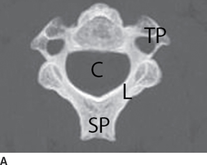
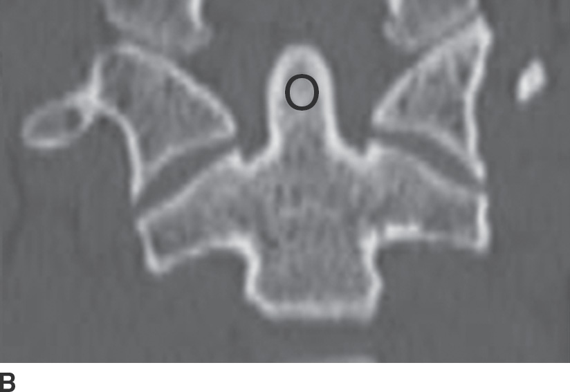
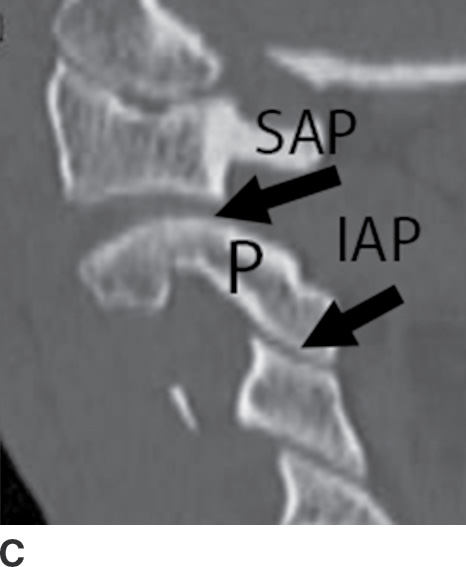
FIG. 10.4 Axial maximal intensity projection (MIP) (A), coronal (B), and sagittal (C) reformatted images of the cervical spine demonstrating the superiorly projecting odontoid process (O), pedicles (P), lamina (L), and the spinal canal (C) are also noted. Note the rounded shape of the superior articular process (SAP) and the more “normal” appearance of the inferior articular process (IAP) on the sagittal MIP. The transverse processes (TP) end in a single tubercle. The spinous process (SP) is bifid in nature.
The C3 to C7 vertebral bodies are wider in the transverse diameter compared to the anteroposterior diameter with a concave upper surface forming the paired uncinate processes. The uncinate processes articulate with the adjacent superior vertebral body’s inferolateral surface to form the uncovertebral joints, located at the posterior and lateral margin on the intervertebral disk. The superior and inferior articular processes are fused, forming the articular pillars. The articular processes are flattened and oval in shape. The vertebral foramen is triangular in shape, formed by the pedicles and lamina posterolaterally. The posterior directed spinous process is usually bifid from C3 to C6 (1,4) (Fig. 10.5).
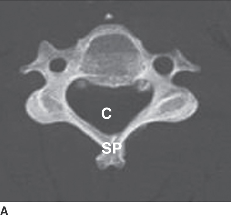
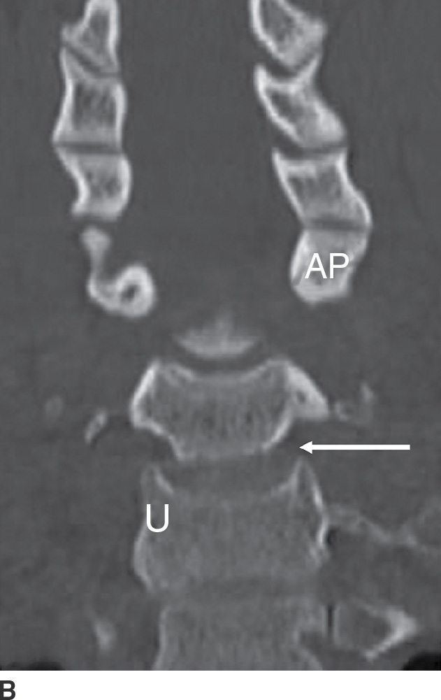
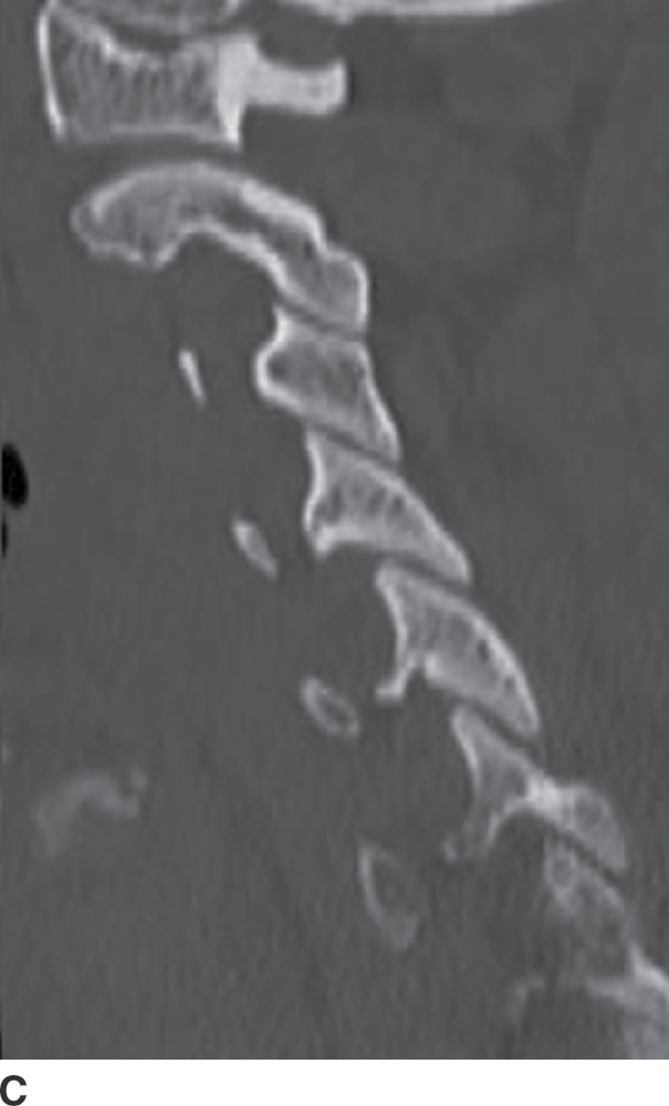
FIG. 10.5 Axial maximal intensity projection (MIP) (A), coronal (B), and sagittal (C) reformatted images of the cervical spine inferior to C2 demonstrating the wide nature of the vertebral body. The paired uncinate processes (U) are seen articulating with the adjacent vertebral body, forming the uncovertebral joint (arrow). The superior and inferior articular processes form the articular pillars (AP). Notice the triangular appearance of the spinal canal (C). The spinous process (SP) is usually bifid from C3 to C6.
The first through sixth vertebrae contain the foramen transversarium (see Fig. 10.3), which is demarcated by the anterior and posterior tubercles of the transverse processes, encasing the V2 segment of the vertebral arteries, the vertebral venous plexus, and sympathetic nerves (1,4). In a study by Bruneau et al., a high incidence of variation of the entry level of the V2 vertebral artery segment was observed. Ninety-three percent of vertebral arteries entered the C6 foramen transversarium, but the entry level varied from the C3 to C7 foramen transversarium. In 2.0% of specimens, a medial loop was present medial to the border of the uncovertebral joint or into the intervertebral foramen (5). Reporting vascular variants to referring clinicians is of paramount importance when spinal interventions are planned.
The cervical neural foramina are oriented at 45-degree angles with respect to the coronal plane and about 10 degrees downward with respect to the axial plane. In the neutral position, the cervical spinal nerves are located in the inferior neural foramen, at or below the disk level (6). However, their position and size of the neural foramina may vary depending on neck positioning (7).
Thoracic spine
There are 12 thoracic vertebrae, each consisting of a body, vertebral arches, and seven processes. The superior articular facets arise from the superior articular processes, and inferior articular facets arise from the inferior articular processes (4). The upper and midlevel paired thoracic facet joints are oriented in the coronal plane, while the cervicothoracic joints are oriented in the axial plane and the thoracolumbar joints are oriented in the sagittal plane (Fig. 10.6) (8). This relationship becomes important when contemplating thoracic facet joint injections. The thoracic spinous processes project from the vertebral arches and slope inferiorly.
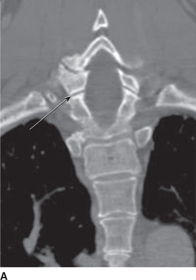
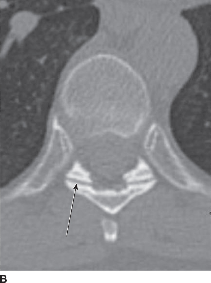
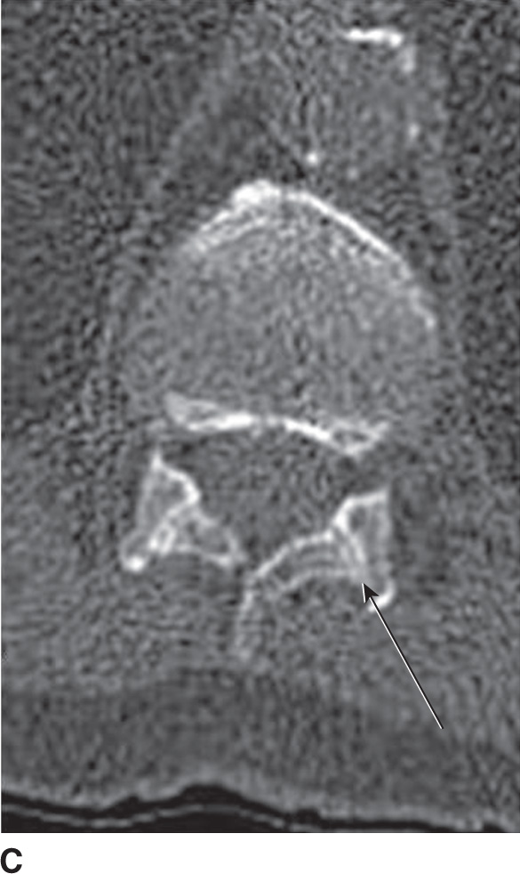
FIG. 10.6 A: Coronal CT of the cervicothoracic spine demonstrating the axial orientation of the zygapophyseal joints (arrow). B: Axial CT of the midthoracic spine demonstrating the coronal orientation of the zygapophyseal joints (arrow). C: Axial CT of the thoracolumbar junction demonstrating the sagittal orientation of the thoracolumbar joints (arrow).
Located at the level of the intervertebral disk, the costal facets articulate with the rib heads. From T2 to T9 levels, these demifacets are paired, where the superior and inferior costal demifacets span two adjacent vertebral bodies (T2 to T9) and articulate with the rib heads. When there are paired demifacets, the more inferior vertebral body identifies its corresponding rib (the third rib articulates with the inferior costal facet of T2 and the superior costal facet of T3) (4). Four thoracic vertebral bodies (T1, T10, T11, T12) have variant anatomy. The superior costal facet of T1 is not a demifacet (i.e., the T1 rib only articulates with T1). The T1 inferior costal facet assumes the usual thoracic anatomy (it is a demifacet). T10, T11, and T12 have paired bilateral facets (no demifacets) (4) (Fig. 10.7).
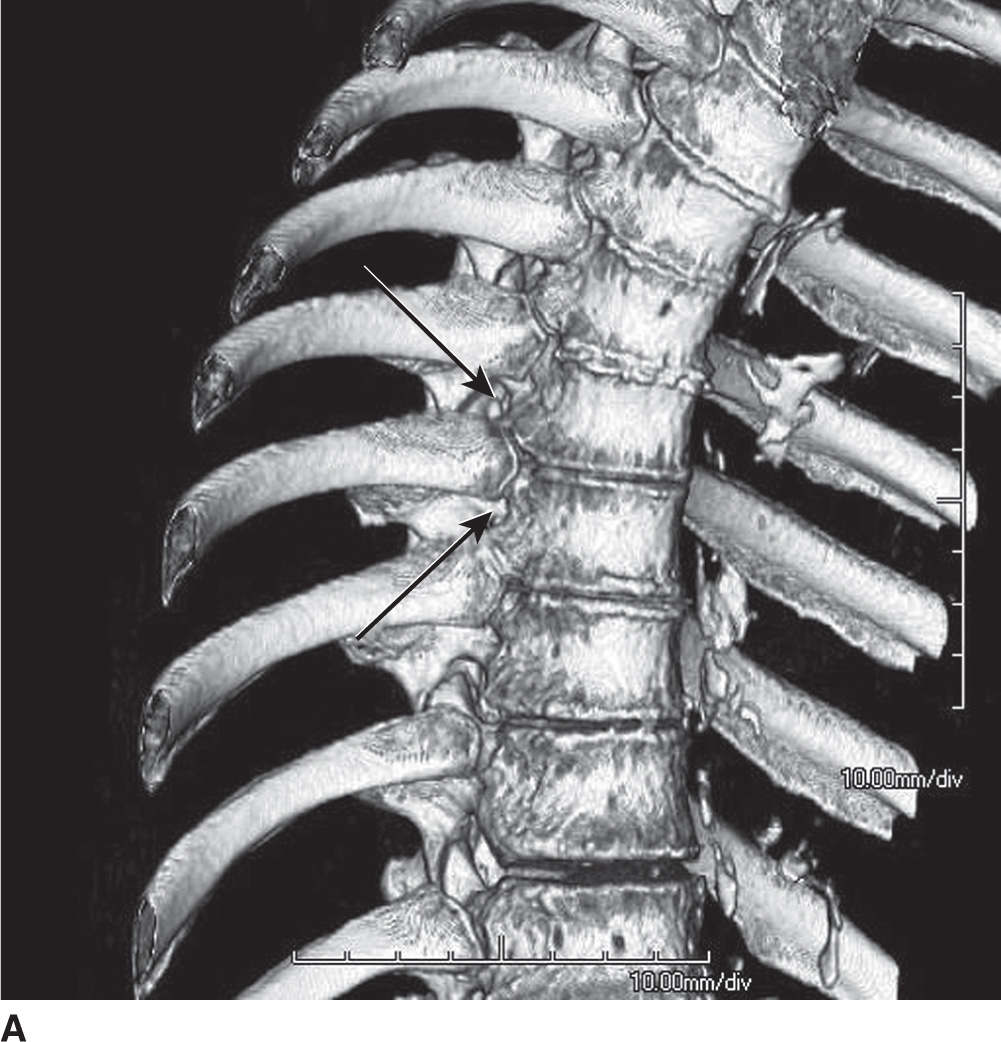
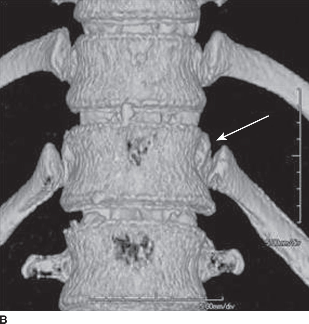
FIG. 10.7 Coronal 3D volume rendered image of the midthoracic spine (A) demonstrating rib articulation with demifacets (arrows) and the lower thoracic spine (B) demonstrating rib articulation with “traditional” facets (arrow).
Lumbar spine
There are five lumbar vertebrae. The lumbar vertebral bodies are thick and wide, with short pedicles and laminae. The inferior articular processes extend inferiorly from the undersurface of each lamina, while the superior articular processes extend superiorly from the junction of the laminae and pedicles. The spinous processes, mamillary processes, accessory processes, and transverse processes serve as muscular attachments and, in the case of the transverse processes and spinous processes, as levers. The zygapophyseal joints are formed by the medial border of the superior articular facet and the lateral border of the inferior articular facet. The pars interarticularis is the portion of the lamina between the superior and inferior articular processes, an area that may experience excessive forces during spinal motion (9) (see Fig. 10.1). The intervertebral or neural foramen is larger than in the thoracic spine, but smaller than in the cervical spine, while the inferior vertebral notches are relatively large (1).
The lateral recess is an area in the lumbar spine bordered laterally by the pedicle, posteriorly by the superior articular facet, and anteriorly by the vertebral body and intervertebral disk. Hypertrophy of the superior articular facet may cause nerve root compression at the superior border of the pedicle (10). The subpedicular notch, located in the anterior and superior portion of the intervertebral foramen, is the usual location of the dorsal root ganglion (DRG) and exiting nerve root, although its location may vary depending on the spinal level (11).
The lumbar intervertebral foramen is a keyhole- or inverted teardrop–shaped area bordered anteriorly by vertebral bodies and intervertebral disk, posteriorly by the ligamentum flavum and superior and inferior articular facets, and superiorly and inferiorly by the adjacent pedicles (11) (Fig. 10.8). The foraminal zone and extraforaminal zone are two locations that radiologists should be familiar with, as they have an increased likelihood of entrapment by disk herniation. The area underneath the pedicle but within the canal is the foraminal zone, and the area outside the lateral border of the pedicle is referred to as the extraforaminal zone (12).
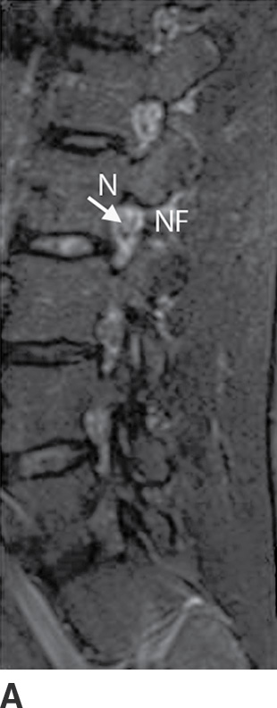
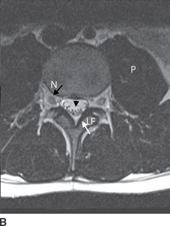

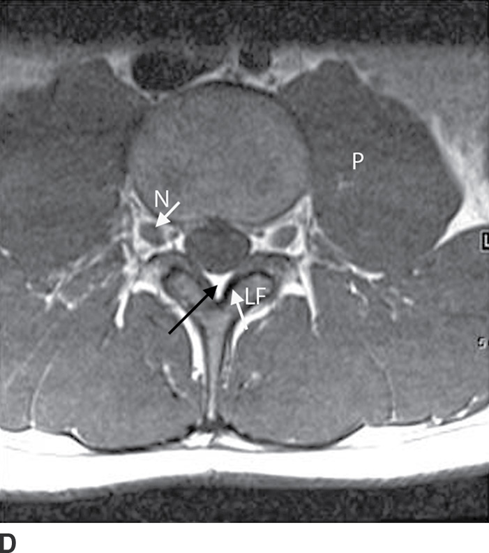
FIG. 10.8 Sagittal T2- (A), axial T2- (B), sagittal T1- (C), and axial T1 (D)-weighted images of the lumbar spine at the level of the neural foramen demonstrate the key hole-shaped appearance of the neural foramen (NF) (A,C) with the exiting nerves (N) in the intervertebral foramen (B,D). Notice the U-shaped appearance of the spinal nerves within the thecal sac (black arrowhead). The ligamentum flavum (LF) is seen as a thick band of decreased signal along the ventral aspect of the lamina. The psoas (P) muscles are also well seen. Epidural fat is present as high signal intensity of T1- and T2-weighted images (black arrow).
Related posts:


Stay updated, free articles. Join our Telegram channel

Full access? Get Clinical Tree


