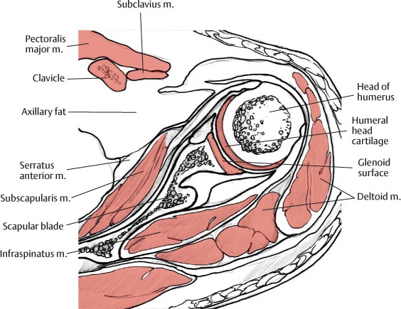
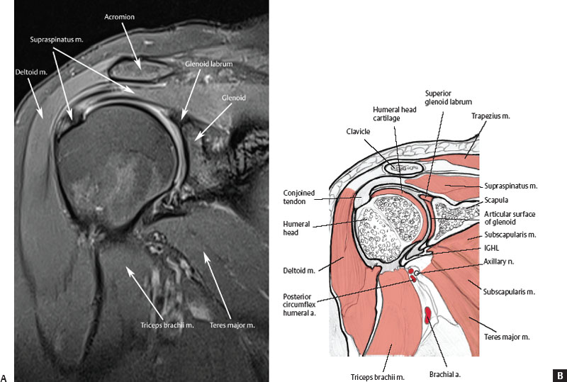
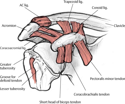
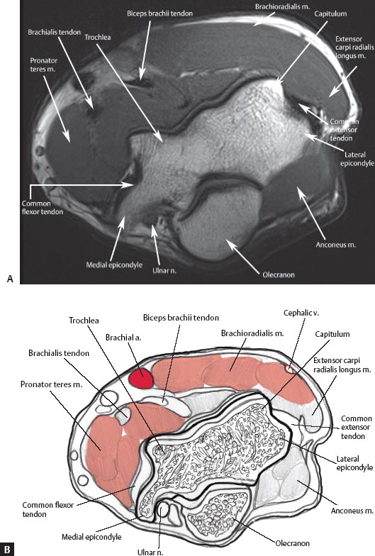
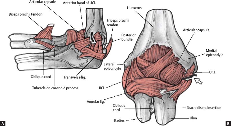
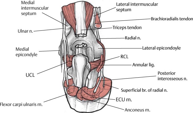
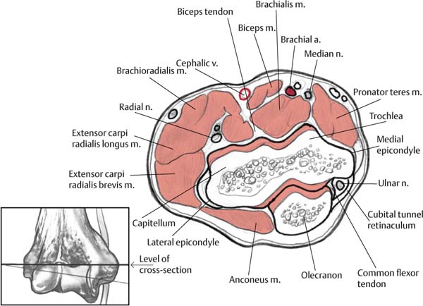
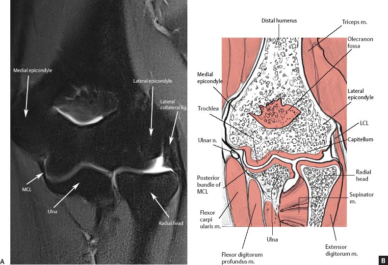
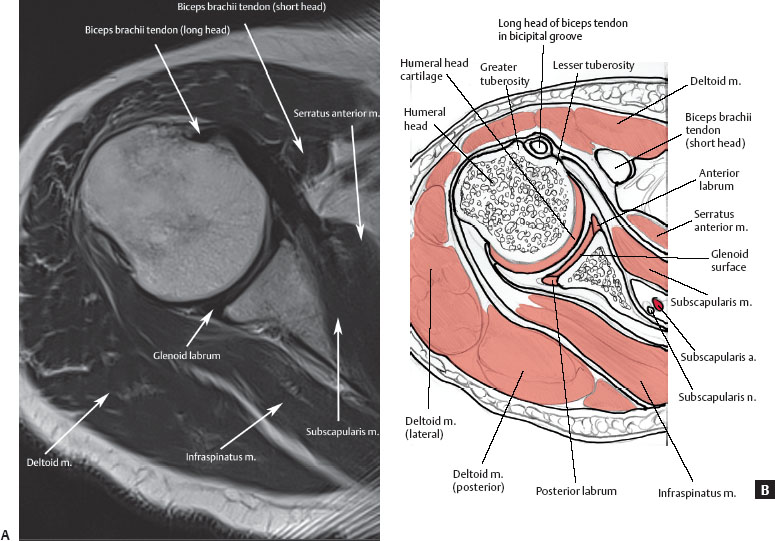
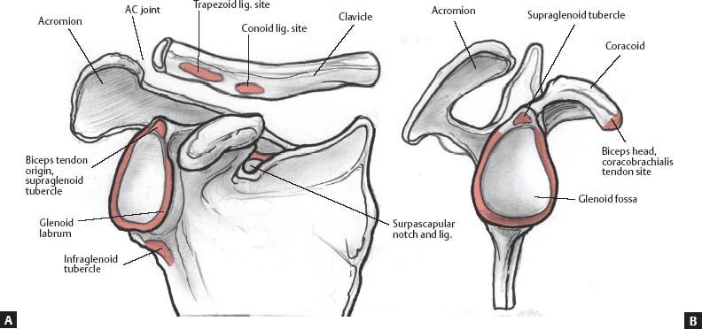
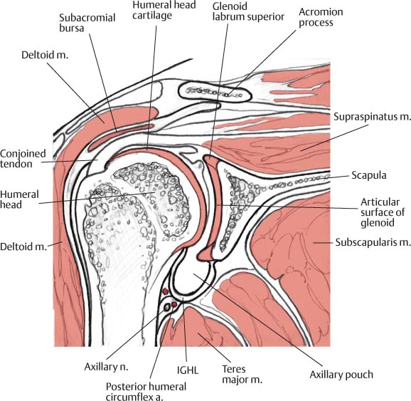
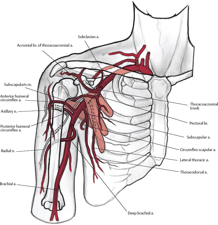
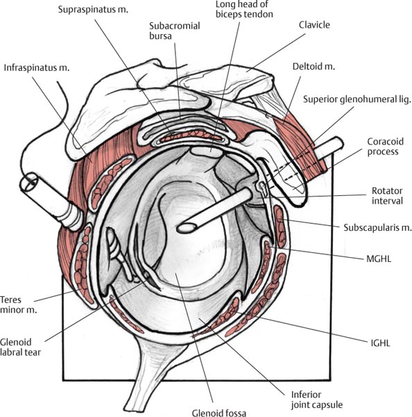
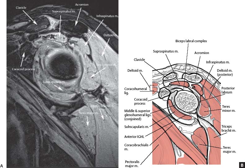
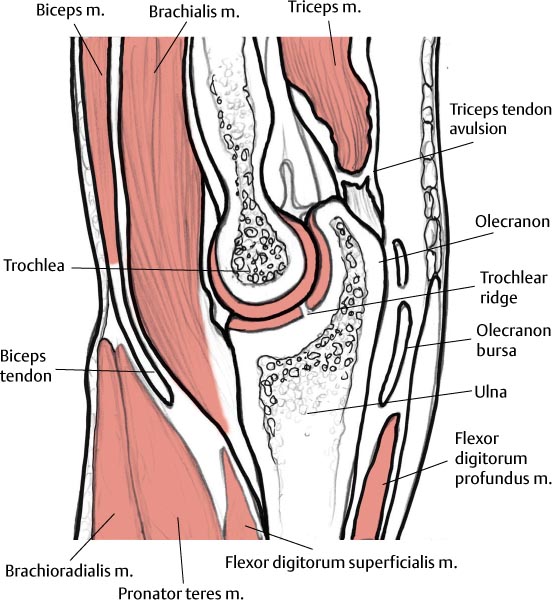
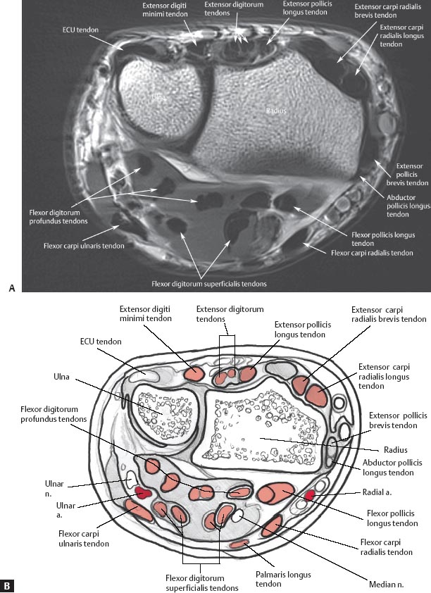
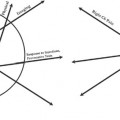
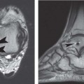
2 Normal MRI Anatomy of the Musculoskeletal System To evaluate effectively an MRI examination of a particular joint or region in the musculoskeletal system, it is essential to have at least a basic understanding of the normal MRI anatomy of that region. Many excellent texts and atlases have been written to serve this need for clinicians and radiologists.1,2 This chapter provides a brief overview of the most clinically important anatomy for each major region of the musculoskeletal system. The figures and line drawings in this chapter serve to highlight the structures with which the musculoskeletal medicine provider should be familiar before interpreting an imaging examination of the region. Reviewing the normal anatomy images pertinent to a specific anatomic region before reading the corresponding region-specific chapter will enhance the clinician’s understanding of the relevant pathologic conditions and facilitate recognition and differentiation of the subtle regional anatomic alternations that represent various pathologic conditions. Axial images are obtained from the superior aspect of the AC joint through the inferior glenoid margin. Axial plane images are best used for evaluating the glenoid labrum (anterior and posterior portions) (Fig. 2.1) and capsular structures as well as the long head of the biceps tendon in the bicipital groove.3 In addition, these images provide good visualization of the subscapularis muscle and tendon, the humeral head, and the glenoid (Fig. 2.2). On superior axial images, the normal oblique course of the supraspinatus muscle is displayed with intermediate signal intensity, and the supraspinatus tendon is low in signal intensity. In cross-section, the tendon of the long head of the biceps is seen as a low signal intensity structure within the bicipital groove. Glenoid articular cartilage follows the concave shape of the glenoid cavity and shows intermediate signal intensity on T1-weighted and T2-weighted images. Articular cartilage of the glenohumeral joint is best evaluated on gradient-echo or fat-suppressed T2-weighted sequences.4–6 The glenohumeral ligaments, best visualized on axial images, have low signal intensity on all pulse sequences. The superior glenohumeral ligament is identified at the level of the coracoid and the biceps tendon. The MGHL is highly variable and may be identified as a thin band or a cord between the anterior labrum and subscapularis or may not be visualized at all without capsular distention. The anterior band of the IGHL is visualized more inferiorly between the anterior inferior labrum and the subscapularis tendon. Coronal oblique images are obtained in a plane that is parallel to the course of the supraspinatus tendon; they tend to show shoulder anatomy and pathology in a plane that is familiar to most. The osseous structures of the shoulder are easily recognized (Fig. 2.3A) as they would be seen on an AP shoulder radiograph. Similarly, the sagittal oblique images show the osseous structures as they would be seen from a lateral view (Fig. 2.3B). The coronal oblique images should include the subscapularis muscle anteriorly and the infraspinatus and teres minor muscles posteriorly. Coronal oblique images are best used to evaluate the supraspinatus muscle and tendon (Fig. 2.4), the subacromial and subdeltoid bursa, and the AC joint. The long head of the biceps tendon and biceps attachment, the infraspinatus muscle and tendon, the glenoid labrum (superior and inferior portions), and the glenohumeral joint space can also be visualized in the coronal oblique plane. Each coronal oblique image should be evaluated systematically from anterior to posterior. On anterior coronal oblique images, the subscapularis muscle and tendon can be identified as the tendon courses from its origin in the subscapularis fossa to its insertion on the lesser tuberosity. However, the subscapularis muscle and tendon can be seen more clearly on axial images (Fig. 2.2). The long head of the biceps tendon is best seen in its intraarticular location on coronal oblique images. On anterior and midcoronal oblique images, the supraspinatus muscle and tendon are seen in continuity (Fig. 2.5). The supraspinatus originates in the supraspinatus fossa of the scapula and inserts on the superior facet of the greater tuberosity of the humerus. On coronal oblique images, the anatomy of the AC joint is best displayed at the level of the supraspinatus tendon. The AC joint should be evaluated for the shape of the acromion (Fig. 2.4) and the various ligaments around the shoulder (Fig. 2.6). The superior and inferior portions of the glenoid labrum, as well as the axillary pouch, are also clearly shown on coronal oblique images. The superior and inferior glenoid labrum are seen as low signal intensity structures, in contrast to high signal intensity fluid on T2-weighted images. The axillary pouch usually is collapsed or has a small amount of fluid in the recess (Fig. 2.5). Humeral head articular cartilage, intermediate in signal intensity on T1-weighted and T2-weighted images, is interposed between the low signal intensity supraspinatus tendon superiorly and the cortex inferiorly. Fig. 2.1 An axial T2-weighted image (A) and artist’s sketch (B) of the right shoulder at the level of the glenoid labrum showing the long head of the biceps tendon as it courses along the bicipital groove. Fig. 2.2 An axial illustration of the left shoulder showing the anterior position of the subscapularis muscle and the articular cartilage of the glenoid and humeral head. Fig. 2.3 Anterior (A) and lateral (B) 3D illustrations of the osseous structures of the right shoulder. Fig. 2.4 A coronal proton-density fat-suppressed image (A) and artist’s sketch (B) of the right shoulder at the level of the supraspinatus muscle and the insertion of the conjoined tendon, a site that is very prone to rotator cuff injury. The normal, flat acromion is also seen at this level, without evidence of supraspinatus impingement. Fig. 2.5 A coronal illustration of the right shoulder at the level of the supraspinatus muscle and tendon showing the glenoid and humeral head cartilage and the superior and inferior glenoid labrum. The subclavian artery courses laterally between the anterior scalene and middle scalene muscles. The axillary artery continues from the subclavian artery at the lateral border of the first rib. Branches of the axillary artery are the supreme thoracic artery, thoracoacromial artery, lateral thoracic artery, subscapular artery, and anterior and posterior humeral circumflex arteries. The brachial artery continues from the axillary artery at the lateral border of the teres major muscle (Fig. 2.7). The brachial artery passes posterior to the bicipital aponeurosis, and its branches provide arterial flow to the forearm and hand. Fig. 2.6 A 3D coronal illustration of the right shoulder, identifying the insertion locations of the rotator cuff tendons at the greater and lesser tuberosities of the humerus. Also shown are the coracoclavicular and coracoacromial ligaments and other ligaments that stabilize the shoulder. Sagittal oblique images, obtained in a plane that is perpendicular to the supraspinatus tendon, should extend from the most lateral aspect of the humeral head to the midscapula (to evaluate rotator cuff muscle atrophy). The osseous structures (Fig. 2.3B) can be used to orient oneself to the location of the rotator cuff muscles, tendons, and other nonosseous structures (Fig. 2.8). These oblique images are well suited for evaluating the rotator cuff muscles and tendons (Fig. 2.9), coracoacromial arch, rotator interval, and acromial morphology.3 The glenoid labrum and the long head of the biceps can also be evaluated on the sagittal oblique images. However, both structures are better visualized on axial and coronal oblique images. Sagittal oblique images should be reviewed systematically from medial to lateral. Medial sagittal sections display the clavicle and AC joint in profile. On midsagittal and lateral sagittal images, the supraspinatus, the infraspinatus, and the confluence of the cuff tendons are visualized between the acromion and the superior articular surface of the humeral head. The supraspinatus originates from the supraspinatus fossa of the scapula, and the infraspinatus originates from the infraspinatus fossa of the scapula. The teres minor originates from the posterolateral aspect of the scapula. All three of these rotator cuff structures (the supraspinatus, infraspinatus, and teres minor) insert at the greater tuberosity of the humerus: the supraspinatus, along the most superior aspect of the greater tuberosity; the infraspinatus, along the middle facet of the greater tuberosity; and the teres minor, along the inferior facet of the greater tuberosity. The subscapularis is the most anterior rotator cuff muscle, and it originates from the subscapularis fossa of the scapula. It is unique in that it is the only rotator cuff structure to insert along the lesser tuberosity of the humerus rather than the greater tuberosity (Fig. 2.6). The biceps tendon can be followed from medial to lateral as it courses from its intraarticular origin within the synovial sheath to its more lateral extracapsular location in the bicipital groove. The long head of the biceps originates from the supraglenoid tubercle, and the short head of the biceps originates from the coracoid (Fig. 2.3). Fig. 2.7 A 3D coronal illustration of the neurovascular structures of the right shoulder and arm showing the subclavian, axillary, and brachial arteries, as well as smaller branch vessels such as the anterior humeral circumflex artery, a tributary of the axillary artery, which is seen coursing anterior to the surgical neck of the humerus. Fig. 2.8 A 3D sagittal illustration of the right glenoid showing a labral tear. Axial images of the elbow should extend from above the humeral epicondyles (Fig. 2.10) to a level distal to the radial tuberosity. The tendons related to the elbow are best evaluated in the axial plane. The major muscles in the anterior compartment of the arm are the biceps brachii and the brachialis; the major muscle in the posterior compartment is the triceps brachii. Ventrally, the biceps tendon is seen as a low signal intensity structure, which courses from its musculotendinous junction, beneath the lacertus fibrosis, to its insertion on the radial tuberosity (Fig. 2.11). Some fibers of the distal biceps brachii also contribute to the bicipital apo-neurosis. The aponeurosis extends from the myotendinous junction of the biceps to the fascia overlying the anteromedial muscles (flexors and pronators) and is identified as a thin, black, low signal intensity line on an axial image. The biceps brachii spans the shoulder and elbow joints and has a short and long head. The brachialis originates from the anterior aspect of the distal humerus, and its tendon courses immediately deep and slightly medial to the biceps and inserts on the ventral surface of the coronoid process of the ulna. The brachialis muscle is intermediate in signal intensity. Posteriorly, the triceps brachii has three heads with three separate origins; the distal triceps tendon attaches to the olecranon process of the ulna (Figs. 2.12 and 2.13). Fig. 2.9 A sagittal proton-density image with fat suppression (A) and artist’s sketch (B) of the left shoulder at the level of the rotator cuff muscles. Although it takes some practice to evaluate the shoulder in the sagittal plane, the rotator cuff muscles are often best visualized on this view. Axial images also show the muscle architecture well. Because of the relative complexity of the forearm musculature compared with the arm musculature, forearm muscles are often grouped by location (superficial or deep) or by compartment (anterior, lateral, or posterior). Both classification schemes are acceptable, although individual radiologists or clinicians may have a preference; it may be helpful to review both classification schemes. There are seven superficial muscles within the dorsal aspect of the proximal forearm: • Extensor carpi radialis brevis • Extensor carpi radialis longus • Brachioradialis • Extensor digitorum • Extensor digiti minimi • ECU • Anconeus (not always present) Five superficial muscles are found within the volar aspect of the proximal forearm: • Flexor carpi radialis • Flexor carpi ulnaris • Flexor digitorum superficialis • Palmaris longus (absent in approximately 15% of the population7) In the proximal forearm, there is only one superficial muscle within the dorsal aspect, the supinator, and one deep muscle within the volar aspect, the flexor digitorum profundus (for the superficial and deep muscles within the distal forearm, see Wrist, below). The anterior compartment of the forearm contains the following five muscles: • Pronator teres • Flexor carpi radialis Fig. 2.10 An axial proton-density image (A) and artist’s sketch (B) of the left elbow at the level of the humeral epicondyles illustrating the muscle architecture of the distal arm. The biceps brachii tendon, the common flexor tendon, and the common extensor tendon show normal thickness and low signal intensity. • Flexor digitorum superficialis • Flexor carpi ulnaris • Flexor digitorum profundus There are four muscles in the lateral compartment of the forearm: • Brachioradialis • Extensor carpi radialis longus • Extensor carpi radialis brevis • Extensor digitorum Two muscles are found in the posterior compartment of the forearm: • Anconeus • ECU Fig. 2.11 Lateral (ulnar side) (A) and posterior (B) 3D illustrations of the left elbow showing the ligamentous structures that stabilize the elbow joint and a tear of the UCL (arrow on B). Fig. 2.12 A posterior 3D illustration of the right elbow showing the insertion of the triceps tendon (cut) onto the olecranon process of the ulna. Also seen are other neural and ligamentous structures. Fig. 2.13 A sagittal illustration of the elbow at the ulnotroch-lear articulation showing an avulsion of the distal triceps tendon near its insertion. Fig. 2.14 An axial illustration of the elbow at the level of the medial and lateral epicondyles (see inset) showing the radial and ulnar nerves and the common flexor and common extensor tendons. Fig. 2.15 An anterior 3D illustration of the neurovascular structures about the right elbow. It is helpful to identify the origin and insertion sites of forearm tendons that involve the elbow because these locations are often the sites of tears and avulsions. Medially, the common flexor tendon is seen on axial images in cross-section as an ovoid, low signal intensity structure originating from the medial epicondyle of the humerus (Fig. 2.14). It is shared by several superficial flexor muscles in the forearm: • Pronator teres • Flexor carpi radialis • Palmaris longus • Flexor digitorum superficialis • Flexor carpi ulnaris Not surprisingly, given their shared origin at the medial epicondyle via the common flexor tendon, the pronator teres and flexor carpi radialis are both involved in medial epicondylitis. The medial epicondyle is not only the origin of the common flexor tendon, but also the origin of the MCL (or UCL). The common flexor tendon is superficial to the MCL, which also attaches to the sublime tubercle, the most proximal and medial portion of the ulna. The sublime tubercle is a helpful landmark, which can be used to easily identify the MCL, especially given that the common flexor tendon and the MCL are similarly low in signal intensity. The LUCL and RCL are low signal intensity structures originating along the lateral epicondyle in the lateral compartment of the elbow (Fig. 2.11). The common extensor tendon is located more posteriorly but also attaches to the lateral epicondyle (Fig. 2.14). It is seen in cross-section on axial images and is low in signal intensity. The common extensor tendon is shared by several extensor muscles in the forearm, including the following: • Extensor digitorum • Extensor carpi radialis brevis • ECU • Extensor digiti minimi The anconeus is fairly isolated from other musculature, located posterolateral to the elbow. Normal muscles are usually intermediate in signal intensity, so the isolation of the anconeus from other structures with similar signal intensity makes it more easily identifiable. The anconeus is a short muscle, spanning only the elbow joint, with its origin at the lateral epicondyle and its insertion along the proximal ulna. The anconeus is a weak extensor, but it also functions to tighten the joint capsule. The supinator is also seen in this general region, although it originates directly from the lateral epicondyle and the olecranon, rather than attaching to the lateral epicondyle via the common extensor tendon. The supinator runs along the radius laterally and inserts along the lateral aspect of the proximal radius. The axial plane allows for evaluation of the neurovascular structures (Fig. 2.15). The median nerve, which lies in close association with the pronator teres and flexor carpi radialis, can often be identified as a thin, intermediate signal structure located between the ulnar and humeral heads of the pronator teres. The brachial artery, seen in close association with the origin of the pronator teres, lies lateral to the median nerve. The ulnar nerve is well visualized posterior to the medial epicondyle within the cubital tunnel (Fig. 2.14), just deep to the cubital tunnel retinaculum, where it is surrounded by high-signal fat on non–fat-suppressed images. The ulnar nerve is normally intermediate in signal intensity. Although not present in most individuals, the anconeus epitrochlearis muscle is a normal variant structure that may overlie the cubital tunnel and ulnar nerve, extending from the posterior aspect of the medial epicondyle to the medial aspect of the ulna. Proximal to the elbow, the ulnar nerve is located along the medial border of the triceps muscle. Distal to the elbow, the ulnar nerve is seen between the two heads of the flexor carpi ulnaris muscle. The radial nerve runs between the brachioradialis and brachialis muscles. At the level of the biceps myotendinous junction, the radial nerve can be seen dividing into its superficial and deep branches within the radial tunnel (Fig. 2.12). The posterior interosseous nerve (deep branch of the radial nerve) is located between the two origins of the supinator. The median nerve lies centrally but is difficult to distinguish from the adjacent brachial vessels. Coronal images of the elbow are obtained in a plane parallel to the interepicondylar line. Although the common flexor and common extensor tendons are seen on axial images, these structures are also well visualized in the coronal plane. The collateral ligament complexes (Fig. 2.16) are best evaluated on coronal images. Starting with the medial aspect of the elbow, the common flexor tendon is seen as it arises from the medial epicondyle. Just deep to this tendon, the anterior band of the UCL courses from the distal margin of the medial epicondyle to its attachment on the sublime tubercle of the proximal ulna. The posterior and oblique bands of the UCL are generally not evaluated well on MRI examinations. The ulnar nerve can be identified posterior to the medial epicondyle and is shown in long axis on the coronal images, allowing for large segments of the ulnar nerve to be visualized on a single image. When evaluating the lateral aspect of the elbow, the common extensor tendon can be identified arising from the lateral epicondyle of the humerus. The RCL lies deep to the common extensor tendon and extends from the anterior aspect of the lateral epicondyle to the radial head, where it inserts onto the annular ligament. The LUCL is also deep to the common flexor tendon but is more posteriorly located relative to the RCL; the LUCL extends from the tubercle of the supinator crest of the ulna to the postero-lateral aspect of the radius. The LCL complex comprises the following: Fig. 2.16 A coronal proton-density fat-suppressed image (A) and artist’s sketch (B) of the left elbow showing the MCL and LCL in the long axis at the level of the radiocapitellar and ulnotrochlear articulations. • LUCL • Annular ligament The articular cartilage of the radiocapitellar and ulnotrochlear joints is well visualized in the coronal plane, but a “pseudodefect” is often identified on the capitellum on more posterior coronal images. This appearance in the coronal plane relates to the fact that the surface of the posterior capitellum is not covered with articular cartilage, often resulting in the appearance of a dark, thin line at the junction of the distal humerus and the capitellum; fluid accumulation in this region may accentuate the appearance of a defect because of its high signal intensity. Awareness of the normal appearance of this pseudodefect is critical: the dark line at the junction of the distal humerus and capitellum could other wise be misinterpreted as an impacted fracture, and the accumulation of fluid in this region could be confused with an OCD. Another area that may mimic a cartilage defect is along the lateral aspect of the radial head, where the articular cartilage is generally thinner than the cartilage in other areas of the elbow. The sagittal plane is useful for evaluating osseous architecture and relationships, especially the humerotrochlear and radio-capitellar articulations (Fig. 2.17). Intraarticular bodies are occasionally difficult to identify because they may have signal characteristics similar to those of joint fluid. The brachialis muscle can be followed to its insertion on the ulnar tuberosity and is seen just anterior to the joint capsule. The biceps muscle can be followed distally to its tendinous insertion on the radial tuberosity. The triceps is best seen on the midline image as it inserts onto the olecranon. Each of these structures can usually be visualized on several images in the sagittal plane. The lateral images show the components of the common extensor tendon originating from the lateral epicondyle, and the medial images show the components of the common flexor tendon originating from the medial epicondyle. In the sagittal plane, the ulnar nerve is often well visualized running posterior to the medial epicondyle. The common flexor tendon lies in close association with the ulnar nerve, and both structures can often be visualized on the same sagittal image. Fig. 2.17 A sagittal T2-weighted image (A) and artist’s sketch (B) of the elbow showing the ulnotrochlear joint and articular cartilage, and the normal appearance of the biceps and triceps muscles in long axis. The normal appearance of the trochlear groove as shown should not be misinterpreted as a cartilaginous defect. The best use of axial images is for evaluating the median and ulnar nerves (Fig. 2.18) and the contents of the carpal tunnel and Guyon canal. Additionally, the flexor and extensor tendons crossing the wrist joint are best visualized in this plane (Fig. 2.19). Many of the muscles in the forearm exert force not only on the elbow but also on the wrist and hand. Muscles in the distal forearm can be classified by location (superficial or deep). Fig. 2.18 Axial illustrations of the musculotendinous units and neurovascular structures of the wrist with retraction of the skin and subcutaneous tissues along the volar (A) and dorsal (B) aspect of the wrist. Fig. 2.19 An axial proton-density image (A) and artist’s sketch (B) of the wrist, proximal to the carpal tunnel, showing the large, low signal intensity musculotendinous units along the volar and dorsal aspects of the left distal forearm. There are five superficial muscles in the dorsal aspect of the distal forearm: • Extensor carpi radialis brevis • Extensor carpi radialis longus • Extensor digitorum • Extensor digiti minimi • ECU Four superficial muscles are found in the distal forearm: • Flexor carpi radialis • Flexor carpi ulnaris • Flexor digitorum superficialis • Palmaris longus (not always present) There are four deep muscles in the dorsal aspect of the distal forearm: • Abductor pollicis longus • Extensor pollicis brevis Fig. 2.20 A 3D illustration of the volar aspect of the dissected right wrist and hand, revealing the neurovascular structures, including the ulnar and median nerves and ulnar and radial arteries. • Extensor indicis Three deep muscles are found in the volar aspect of the distal forearm: • Flexor digitorum profundus • Flexor pollicis longus • Pronator quadratus The flexor carpi radialis originates from the common flexor tendon at the medial epicondyle and inserts on the base of the second and third metacarpals. The muscle belly of the flexor carpi radialis is intermediate in signal intensity, and its tendon should be low in signal intensity. The palmaris longus lies medial to the flexor carpi radialis, originating from the common flexor tendon at the medial epicondyle and inserting along the flexor retinaculum and palmar apo-neurosis. Both the flexor retinaculum and palmar aponeurosis are seen as dark bands within the superficial palmar aspect of the wrist. The flexor carpi ulnaris originates from the common flexor tendon and the olecranon; inserts on the fifth metacarpal, hamate, and pisiform; and lies in close association with the ulnar nerve, which is seen as a thin, intermediate signal intensity structure running just deep and lateral to the flexor carpi ulnaris (Fig. 2.20). The flexor digitorum superficialis originates at the medial epicondyle, coronoid process, and radial head. It inserts along the volar aspect of the middle phalanx of each finger (Fig. 2.21). Proximal to its insertions, the flexor digitorum superficialis divides into four musculotendinous units within the distal forearm, before entering the carpal tunnel. Within the carpal tunnel, the median nerve most often lies immediately superficial to the flexor digitorum superficialis tendons (Figs. 2.18, 2.19, and 2.20), although it may have an intertendinous position. However, within the forearm, the median nerve lies immediately deep to the flexor digitorum superficialis muscle. The large, dark musculotendinous units of the flexor digitorum superficialis are easily identified as they converge within the forearm to enter the carpal tunnel. In contrast to the large, low signal intensity musculotendinous units of the flexor digitorum superficialis, the median nerve is a thinner structure with intermediate signal intensity, which can be more challenging to identify. Thus, the musculotendinous units of the flexor digitorum superficialis may be used as a landmark for the adjacent median nerve. The flexor digitorum profundus (Fig. 2.22) originates from the anteromedial aspect of the ulnar and interosseous membrane and inserts along the distal phalanges of each finger (Fig. 2.21). As does the flexor digitorum superficialis, the flexor digitorum profundus divides into four musculotendinous units within the forearm before entering the carpal tunnel. Within the carpal tunnel, the tendons of the flexor digitorum profundus lie immediately deep to the tendons of the flexor digitorum superficialis and are easy to identify based on this predictable relationship. The flexor pollicis longus originates from the coronoid process of the ulna, the mid-radius, and the interosseous membrane. It inserts along the volar aspect of the base of the distal phalanx of the thumb. The pronator quadratus originates from the volar aspect of the distal ulna and inserts along the volar aspect of the distal radius. The extensor carpi radialis longus originates along the supracondylar ridge of the humerus and the lateral inter-muscular septum. It inserts along the dorsal aspect of the second (index finger) metacarpal base. The extensor carpi radialis brevis originates from the common extensor tendon at the lateral epicondyle and inserts at the dorsal aspect of the third metacarpal base. The extensor digitorum (Fig. 2.18) originates from the common extensor tendon at the lateral epicondyle and inserts along the dorsal aspect of each finger (digits 2 through 5) (Fig. 2.21). The extensor digiti minimi (Fig. 2.23) originates from the common extensor tendon at the lateral epicondyle and inserts along the dorsal aspect of the fifth digit. The extensor digiti minimi runs immediately superficial to the radioulnar joint. The ECU originates from the dorsal aspect of the mid-ulna and from the lateral epicondyle. It inserts at the base of the fifth metacarpal. Proximal to its insertion, it can be seen coursing along the groove of the distal ulna. The pisiform is actually a sesamoid bone within the ECU. The TFCC is composed of the following: • Fibrocartilaginous articular disc • Dorsal and volar radioulnar ligaments • Ulnolunate ligament • Ulnotriquetral ligament • UCL • ECU subsheath • A variable meniscus homologue The abductor pollicis longus originates from the dorsal aspect of the proximal third of the radius and ulna and from the interosseous membrane. It inserts at the dorsal aspect of the first metacarpal base. The extensor pollicis brevis originates at the posterior aspect of the distal third of the radius and the interosseous membrane. It inserts at the dorsal aspect of the base of the proximal phalanx of the thumb, forms the lateral margin of the anatomic snuffbox, and runs in close association with the abductor pollicis longus. These tendons run deep to the extensor retinaculum at the level of the distal radial groove (Fig. 2.23
 Shoulder
Axial Images
Coronal Oblique Images
Sagittal Oblique Images
 Elbow
Elbow
Axial Images
Muscle Classification by Location
Muscle Classification by Compartment
Tendons, Ligaments, and Neurovascular Structures
Coronal Images
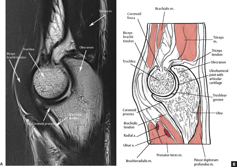
Sagittal Images
 Wrist/Hand
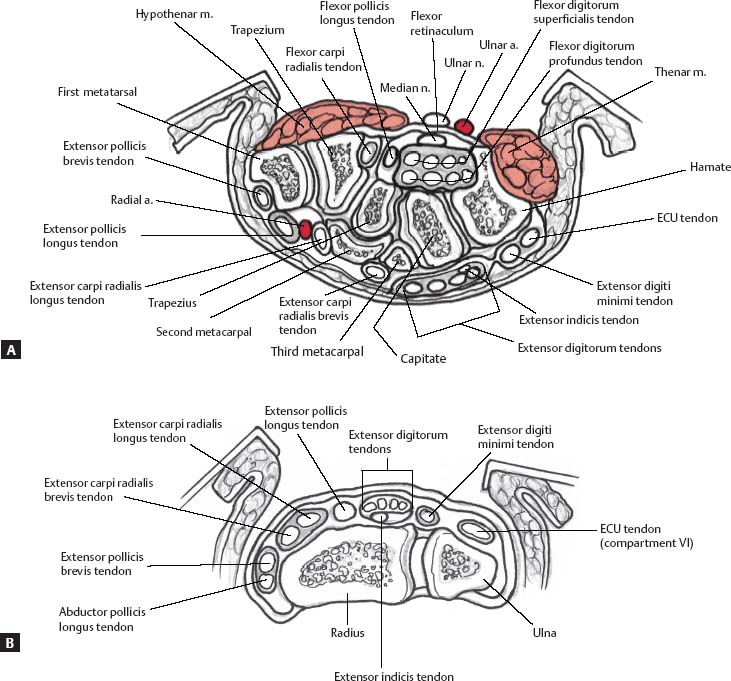
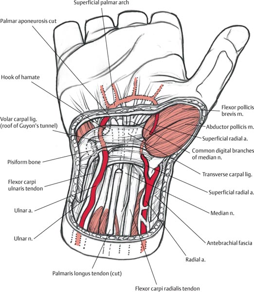
Axial Images
![]()
Stay updated, free articles. Join our Telegram channel


Full access? Get Clinical Tree


