George M. Bridgeforth and George Holmes
A 26-year-old woman presents after falling 4 weeks earlier. She complains of moderate-to-severe pain, swelling, and tenderness of her foot, with pain on movement and difficulty putting weight on it.
CLINICAL POINTS
- Metatarsal fractures are often caused by trauma.
- People with diabetes have a greater risk of infection as a result of a metatarsal injury.
- Stress fractures may manifest as point tenderness, especially over the second or third metatarsal or the fifth metatarsal shaft.
Clinical Presentation
Metatarsal injuries are common, because there is minimal soft tissue to protect the top of the foot. These injuries usually involve direct trauma (impact) but may result from an axial load with torsion. Injuries to the first, second, third, and fourth metatarsals are considered in this chapter, and fractures of the fifth metatarsal fractures are discussed in Chapter 25. Patients with acute metatarsal fractures have moderate-to-marked pain, swelling, and tenderness at the midfoot, which are always important clues that direct the examiner to the fracture site. In addition, patients commonly have pain-limited dorsiflexion and plantar flexion with limited weight bearing.
The clinician should perform a careful neurovascular examination to check the neurovascular status of the foot for evidence of sensory loss, marked swelling, coldness, pallor, and impaired pulses. It is important to document the results. It is necessary to inspect the foot, including each phalanx, carefully for coldness and cyanosis, and palpate the posterior tibial pulse at the posterior medial malleolus just behind the distal tibia. The dorsalis pedis pulse lies medially to the extensor hallucis tendon as it runs along the first metatarsal shaft.
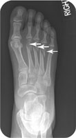
In addition, the clinician should inspect the foot carefully for open wounds. Even with small open wounds without fractures, patients with diabetes are at increased risk for developing a serious cellulitis of the foot (Fig. 26.1). These patients require close follow-up to watch for signs of infection, especially with penetrating wounds, and serious consideration for possible antibiotic therapy with close follow-up. It is also necessary to closely monitor people with diabetes and closed foot injuries for infection (Fig. 26.2).
Patients with multiple metatarsal fractures may have an associated Lisfranc ligament injury. The Lisfranc ligament is at the midfoot and anchors the tarsal bones to the proximal metatarsals. It is important to note that patients with moderate swelling and tenderness over the midfoot may have associated damage to the Lisfranc ligament with or without accompanying metatarsal fractures. In addition, these patients exhibit an antalgic gait (i.e., a pronounced limp). Moreover, they may have trouble wearing regular shoes because of pronounced pain at the dorsal midfoot. Lisfranc injuries may be easily missed, and a high index of suspicion is required (see Chapter 27). The examiner should suspect an associated Lisfranc injury if there is marked tenderness over the Lisfranc (tarsometatarsal) joint (Fig. 26.3).*
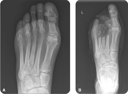
FIGURE 26.1 (A) Notice the lucent areas over the middle metatarsal–phalangeal joints compatible with subcutaneous gas from gas-forming bacteria in left foot cellulitis in this 83-year-old diabetic male. (B) Follow-up radiograph demonstrates midtarsal amputation of the second, third, and fourth digits in the same patient.
Related posts:


Stay updated, free articles. Join our Telegram channel

Full access? Get Clinical Tree


