Fig. 1
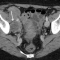
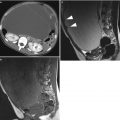
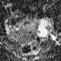
Clear cell carcinoma with paraneoplastic migratory thrombophlebitis in a 40-year-old woman who presented with sudden respiratory distress by pulmonary embolism. (a) Contrast-enhanced CT of the thorax shows pulmonary embolisms in the bilateral pulmonary (arrowheads). (b) CT at the inguinal level shows a venous thrombus in her right femoral vein (arrow). (c) CT of the pelvis demonstrates a well-demarcated cystic mass containing irregular-shaped solid components ventrally (arrowheads). Ascites (asterisk) is present in her left pelvic cavity
CCCs usually present as a large cystic mass, and they rarely occur in bilateral. CCC tends to present in early FIGO stages, with 35–60 % in stage I disease and 9–22 % in stage II. However, stage I CCC is more likely to be stage IC, probably because of higher risk of tumor rupture [6]. Their tendency of early clinical stage at presentation is explained by that CCC usually arises from preexisting endometriomas. However, clear cell carcinoma in early stages had a poorer outcome or a tendency toward poor outcome compared with other ovarian carcinomas [7, 8].
The gross appearances of CCC are characterized by multiple mural nodules within a thick-walled unilocular cyst, which represent endometriomas (Fig. 2) [4]. On MR imaging, the content of the cysts typically shows high intensity on T1-weighted images, as usually seen in endometriomas. However, the signal intensity on T2-weighted images is usually high, rather than low intensity commonly seen in endometriomas [9]. The neoplastic nodule in CCC can show variable size, ranging from tiny nodule to large mass that is about to fulfill the entire cystic cavity. On MR imaging, they usually show low intensity on T1-weigthed images, but variably low to high intensity on T2-weighted images (Fig. 3). Post-contrast image reveals enhancement in the nodules. The confirmation of enhancement on post-contrast image is important for differentiating neoplastic mural nodules from coagulated clots, which are commonly seen in endometriomas but never enhanced. Imaging features in CCCs described above are also seen as endometrioid adenocarcinomas, which can also arise from endometrioma. However, the presence of simultaneous thromboembolitic events, including thrombophlebitis, pulmonary embolisms, and infarctions of many organs, may suggest the diagnosis of CCC (Figs. 1 and 3).
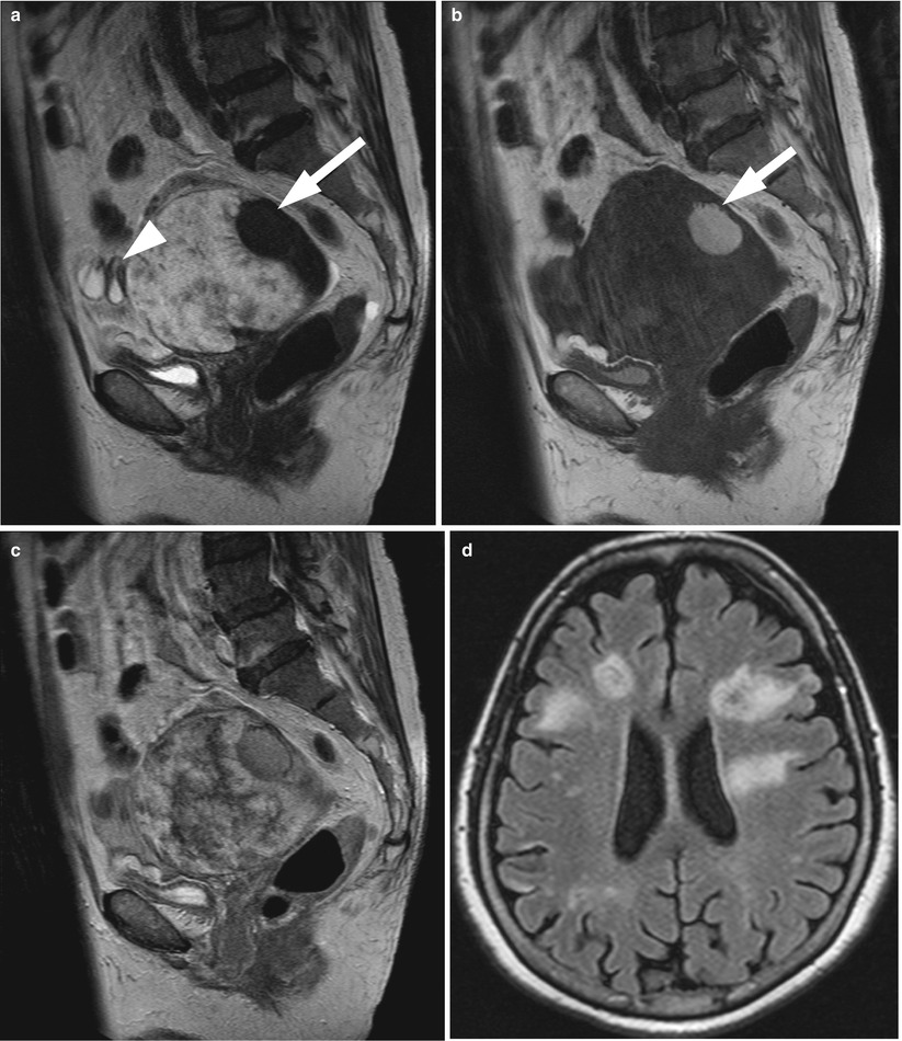
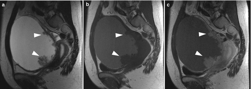

Fig. 2
Clear cell carcinoma in a 64-year-old woman who presented with multifocal acute cerebral infarction. (a) Sagittal T2-weighted image demonstrates an irregular-shaped tumor of high intensity (arrowheads) and endometrioma of distinct low intensity (arrow). (b) T1-weighted image shows predominantly low intensity in the tumor and high intensity in the endometrioma (arrow). (c) Contrast-enhanced T1-weigthed image shows heterogeneous enhancement in the tumor. (d) Fluid attenuated inversion recovery (FLAIR) image shows multiple foci of high intensity, representing acute embolitic infarctions

Fig. 3
Clear cell carcinoma in a 57-year-old woman. (a) Sagittal T2-weighted image shows a large unilocular cyst, associated with multiple papillary projections of intermediate intensity (arrowheads). (b) T1-weighted image shows increased signal intensity in the cyst content and mural nodules of low intensity. (c) Post-contrast T1-weighted image shows enhancement in the papillary projections (arrowheads)
Clear Cell Adenofibromas/Clear Cell Adenofibromas of Borderline Malignancy
Clear cell adenofibromas (CCAs) of borderline malignancy is also called as atypical proliferative clear cell tumor [4]. The gross appearance of these tumor is characterized by microcystic structures of honeycomb appearance, embedded within firm rubbery stroma [4]. Histology of these tumors shows tubular glands lined by a layer of hobnail cells. In CCA of borderline malignancy, the glands are more crowded, and the cells display nuclear atypia. These tumors are considered to be precursors of CCCs, and clear cell carcinoma may accompany these tumors. As like CCCs, these tumors may associate with endometrioma. Although imaging findings of CCAs have not been documented, our experience case of CCA appeared as multilocular cystic mass containing numerous fine septi (Figs. 4 and 5). The areas with abundant fibrous stromas in CCA were demonstrated as solid component of low intensity on T2-weighted images (Fig. 4).
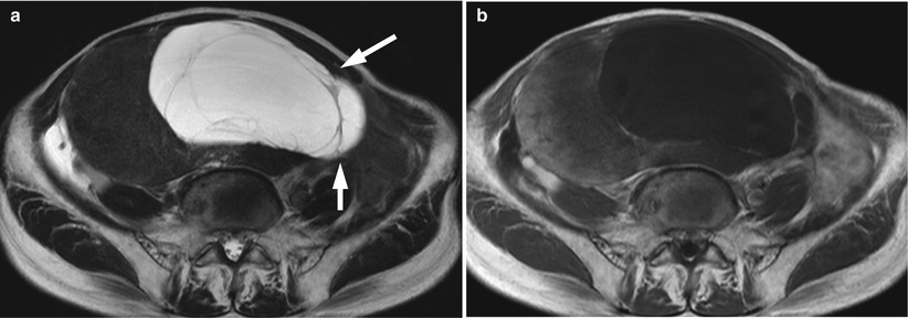
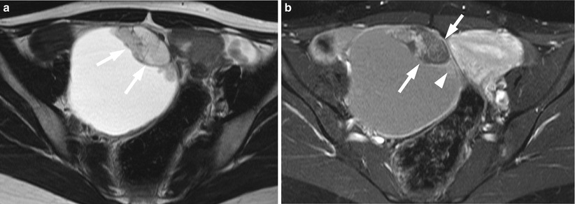

Fig. 4
Clear cell adenofibroma in an 89-year-old woman. (a) Axial T2-weighted image shows a large solid mass containing a cystic component of multilocular appearance (arrows) with fine septi. (b) Post-contrast T1-weighted image shows week enhancement in the periphery of the solid component

Fig. 5
Clear cell borderline cystadenofibroma in endometrioma in a 32-year-old woman. (a) Axial T2-weighted image shows complex mural nodules of heterogeneous intermediate intensity (arrows). (b) Post-contrast-enhanced T1-weighted image reveals microcystic structure within mural nodules with numerous fine septi (arrows). The papillary projections (arrowhead) beside the microcystic lesion are enhanced. These projections correspond to clear cell carcinomas arising from clear cell adenofibroma
Malignant Transitional Cell Tumor
Transitional cell tumors comprise approximately 10 % of ovarian epithelial tumors, and nearly all of these are benign Brenner tumors. Borderline Brenner tumor and malignant transitional cell tumor are very uncommon. Borderline Brenner tumor is also called as atypical proliferative Brenner tumor, proliferating Brenner tumor or Brenner tumor of low malignant potential [4]. Malignant transitional cell tumors are further subdivided into malignant Brenner tumor and transitional cell carcinoma (TCC). These tumors are characterized by the histologic stromal invasion by transitional tumor cells, while this finding is absent in borderline tumor.
Malignant Brenner tumor is pathologically defined by the presence of benign Brenner components within the tumor, while transitional cell carcinoma does not contain these components. Recent reports on immunohistochemical and genetic studies have shown that Brenner tumor has true urothelial differentiation, while TCC is a variant of high-grade serous carcinoma [10, 11]. This recognition is also supported by the observation that high-grade serous adenocarcinomas frequently have focal areas that display features like of TCC [4].
Both borderline and malignant Brenner tumors are usually larger than their benign counterparts and tend to be multilocular cystic in appearance (Figs. 6 and 7). The tumors typically contain multiple papillary projections and irregular-shaped solid components (Fig. 7) [12]. Meanwhile, the gross appearance of TCC is similar to high-grade serous carcinomas and typically is an irregular-shaped solid mass. On T2-weighted images of MR imaging, the signal intensity of solid components have been reported to be higher in malignant transitional cell tumors compared with those in benign and borderline Brenner tumors [13].
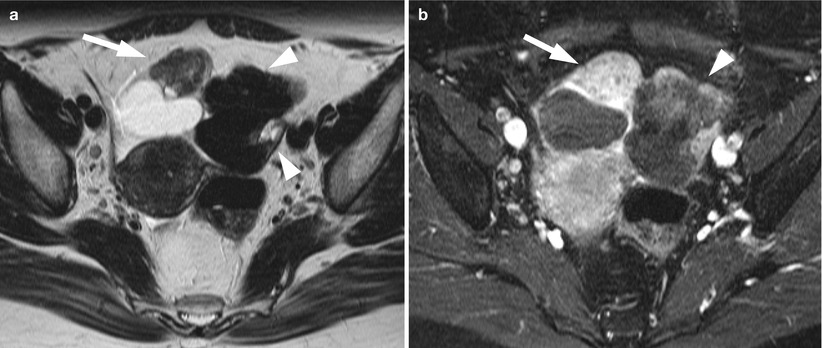

Fig. 6


Borderline Brenner tumor in the right ovary and benign Brenner tumor in the left ovary. (a) Axial T2-weighted image shows bilateral ovarian tumors. Right ovarian tumor is composed of a cystic component dorsally and a solid component (arrow), which exhibits heterogeneously intermediate intensity. Left ovarian tumor is a lobulated solid mass of distinct low intensity (arrowheads). (b) Post-contrast T1-weighted image with fat suppression demonstrates marked but heterogeneous enhancement in the solid component (arrow) of the right ovarian mass and poor enhancement in the left ovarian mass (arrowhead)
Stay updated, free articles. Join our Telegram channel

Full access? Get Clinical Tree


