George M. Bridgeforth, Kathryn J. Mccarthy, and Charles Carroll IV
A 36-year-old man who fell from a ladder presents with right wrist pain.
CLINICAL POINTS
- Pronounced subluxation or dislocation results in a palpable deformity.
- Vascular damage may occur.
- Long-standing cases may lead to degenerative arthritis due to carpal instability.
Clinical Presentation
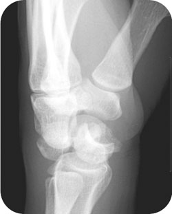
Perilunate dislocation occurs when the lunate remains in a normal position with respect to the distal radius while other carpal bones are dislocated posteriorly. The lunate may be forced into the carpal canal. A relatively rare injury, a perilunate dislocation is commonly associated with scaphoid and transscaphoid wrist fractures. The most common mechanism of injury is a fall onto an outstretched hand. Motor vehicle accidents can also cause volar dislocations of the lunate (Fig. 42.1).
In addition, the volar skin can become ischemic as a result of pressure from the volar radius. Patients may present with arterial compromise or compartment syndrome. As with any acute traumatic injury of the wrist, it is important to check and document the neurovascular status carefully (coldness, pallor, diminished or absent pulses, sensory impairment).
Radiographic Evaluation
It is necessary to order posteroanterior (PA), lateral (Fig. 42.2), and oblique radiographs.
With a perilunate dislocation, the examiner should look for an overlapping of the dorsal displaced capitate, which partially overlies the volar subluxed lunate (PA view). The space between the capitate and lunate is obliterated. The overlapping region has a triangular configuration that is known as a “piece of pie” sign.
Normally, on the lateral view, the alignment of the radius, lunate, and capitate should be within 10 degrees and is shaped like three sequential “C”s (Fig. 43.3). The examiner can think of it as three “C”s in a row. If the middle “C” is displaced volarly, the examiner should suspect a perilunate dislocation. With a lunate dislocation, there is a volar subluxation of the middle “C” (the lunate). The subluxed lunate resembles a tipped teacup that is spilling tea onto the palm. The three “C”s are the concave surface of the distal radius, the lunate (the middle “C”), and the concave surface of the capitate. There is a gap between the radius and capitate (in that gap lies the volar displaced lunate).
If there is a volar subluxation of the lunate (spilled teacup sign), the condition is a lunate dislocation (Fig. 42.3). In these cases, the lunate shows volar displacement beneath a horizontal line constructed through the long axis of the radius and through the middle of the articulating lunate and capitate. Essentially, the middle “C” (the concave surface of the lunate) is beneath this axis, tipped over and pointed downward toward the palm. This finding is best appreciated on the lateral view. However, if there is a dorsal subluxation of the capitate (the third “C”) onto the volar subluxed lunate, so that the capitate subluxation is more pronounced, this condition is referred to as a perilunate dislocation. However, the capitate subluxes dorsally and sits on top of the lunate disrupting the almost straight-line relationship of the three “C”s anatomical disruption may be identified on the lateral view. Although the linear radiolunate axis appears to be maintained (on the lateral view), there may be an associated disruption of the scapholunate ligament.
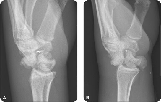
FIGURE 42.1 Various examples of perilunate dislocations. (A) A 26-year-old man with left wrist pain after falling demonstrates volar displacement of the carpal bones (arrow) relative to the normally positioned lunate. (B) A 62-year-old male with unspecified trauma demonstrates a perilunate dislocation. He also sustained a fracture to the scaphoid, not clearly shown.
Related posts:


Stay updated, free articles. Join our Telegram channel

Full access? Get Clinical Tree


