George M. Bridgeforth, Jessica Peelman, and Charles Carroll IV
A 21-year-old man presents with laceration and gross deformity of the fifth finger of his left hand after a motor vehicle collision.
CLINICAL POINTS
- Finger fractures often occur in sports activities
- Without proper treatment, loss of hand function can occur
Clinical Presentation
Phalangeal fractures may occur in the proximal, middle, or distal phalanx, with the distal phalanx being the most commonly fractured bone in the finger. These fractures may lead to a significant loss of hand function if not properly treated. The mechanism of injury is usually direct trauma to the digit when the finger is hit by an object or during a fall. Crushing injuries, twisting injuries, or axial loads (jamming of the finger) may also lead to phalangeal fractures. These injuries are also frequently sports-related, especially in younger patients.
Clinical examination is essential. An important clinical test is to have the patient clinch his or her fist. Normally, the tips of fingers should all point to the scaphoid. If one of the fingers points away from the scaphoid, the examiner should suspect a fracture with a torsion deformity of the finger. It is also necessary to check for tendon injuries by assessing the patient’s ability to independently flex and extend each joint in the finger—the distal and proximal interphalangeal joints as well as the metacarpophalangeal joint. In addition, it is important to check for wounds, nail bed injuries, and open fractures. There may be other associated injuries of the hand or fingers as well. Comparison with fingers of the contralateral hand is crucial. Finally, it is always essential to document the neurovascular status carefully.
PATIENT ASSESSMENT 
- Marked tenderness, swelling, and deformity at the site of the fractured phalanx
- Pain-limited range of motion of the digit and grip strength
- Bruising and ecchymosis
Radiographic Evaluation
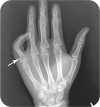
Radiographic tests include posteroanterior (PA), lateral (Fig. 45.1), and oblique views. If the injury is isolated to one finger, finger radiographs provide better resolution. If the injury involves multiple fingers or the hand, hand radiographs are more appropriate.
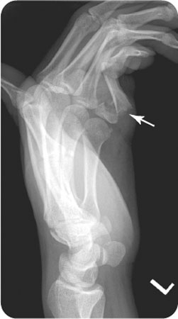
FIGURE 45.1 Lateral radiograph of the left hand from the patient in the introductory case, demonstrating an acute, volar displaced fracture of the fifth proximal phalanx.
Related posts:


Stay updated, free articles. Join our Telegram channel

Full access? Get Clinical Tree


