George M. Bridgeforth and Kris Alden
A 35-YEAR-OLD WOMAN WITH PSORIASIS COMPLAINS ABOUT BILATERAL HAND PAIN AND BACK PAIN.
CLINICAL POINTS
- The large majority of cases of this oligoarticular arthritis are asymmetric.
- Psoriatic sacroiliitis may lead to complaints of low back pain.
- Tendinitis may occur.
Clinical Presentation
Psoriatic arthritis is an oligoarticular, asymmetric arthritis. It may affect the distal interphalangeal (DIP) joints of the fingers, the spine, the sacroiliac joints, the hips, the knees, and the ankles. The diagnosis should be suspected in any patient with an acute joint inflammation and a history of psoriasis, an immune skin disorder. Psoriasis is characterized by salmon-colored plaques with fine silver reticular striae. Common sites of psoriasis include the knees, the tips of the elbows, the gluteal folds, the shins, as well as the soles and palms of the hands.
Psoriatic arthritis affects between 2% and 3% of the general population. It is has an equal distribution among males and females and is more common in Caucasians. The arthritic condition occurs in approximately 5% to 8% of the patients with psoriasis.
In psoriatic arthritis, nail changes, including discoloration, fungal infections (onycholysis of the nail), and small pinpoint depressions (nail pitting) may occur in up to 50% of cases. Yellow–pink and yellow–brown discolorations of the nails (oil-drop sign) may also occur. Tendinitis may be present, and patients may complain of swollen fingers. Such sausage-like swelling of the digits is referred to as dactylitis.
There are five types of psoriatic arthritis, as described below.
- Asymmetric: typically involves three joints or less on different sides of the body and is generally mild.
- Symmetric: usually affects the same joints, in multiple matching pairs, on both sides of the body. This form of disease, which is somewhat similar to rheumatic fever (see Chapter 51, “Rheumatoid Arthritis”), is more severe than the asymmetric type and may be disabling.
- Spondylitis: involves the spine and neck, leading to inflammation and stiffness. It may also affect the joints of the arms, hips, legs, and feet.
- DIP predominant: generally involves the most distal joints in the fingers and toes, resulting in inflammation, stiffness, and nail changes.
- Arthritis mutilans: predominantly affects the small joints in the fingers and toes closest to the nail but often is associated with back and neck pain. This severe form of psoriatic arthritis is deforming and destructive.
PATIENT ASSESSMENT 
- May be associated with pitting of the nails (up to 50% of the cases)
- Early involvement: sausage digits and swollen DIP joints and involvement of the fingers
- Late involvement: pencil-in-cup deformities and acro-osteolysis (flame-shaped erosions of the distal tufts of the hands and feet)
Radiographic Evaluation
Psoriatic arthritis may affect several different joints, including the hands, the feet, and the sacroiliac joints. Radiographic evaluation depends on the affected area. If the sacroiliac joint is affected, radiographs of this joint may be helpful, as well as advanced imaging modalities including computed tomography and/or magnetic resonance imaging.
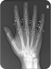
The examiner should look for oligoarticular involvement of the DIP joints of the hands. Early psoriatic involvement of the hands or feet can result in sausage digits. On the radiograph, look for a sausage-like pattern of soft swelling around a digit (Fig. 52.1). In addition, the examiner should observe radiographs for bone erosions at the joint margins of the joints (second and third phalanges) associated with new bone formation at the proximal border. These changes result in a characteristic mouse ear deformity (“Mickey mouse” sign) of the DIP joints (Fig. 52.2). The mouse ear configuration associated with erosive changes is a common feature of advanced psoriatic arthritis. Other advanced changes include the development of tuft erosions of the distal phalanx. This type of terminal erosion, which may appear as a small flame on a candle, is called an acro-osteolysis. Moreover, advanced cases may manifest as an asymmetric ankylosis of the proximal interphalangeal joints and the DIP joints. Other advanced findings of psoriatic arthritis include the development of pencil-in-cup deformities (Fig. 52.3). With this classic deformity, there is a marked erosion of the distal portion of the proximal joint so that it appears like a pencil that is surrounded by a cup.
NOT TO BE MISSED 
- Contusion
- Fracture
- Cellulitis
- Septic joint
- Osteoarthritis
- Osteomyelitis
- Rheumatoid arthritis
- Gout
Related posts:


Stay updated, free articles. Join our Telegram channel

Full access? Get Clinical Tree


