Radiofrequency Pulse Sequences
Objectives
At the completion of this chapter, the student should be able to do the following:
Key Terms
Although the free induction decay (FID) is the primary magnetic resonance imaging (MRI) signal, it is difficult to use in practice. A hard radiofrequency (RF) pulse must be transmitted into the patient at the Larmor frequency to obtain the FID. The FID, which is a weak signal, also at the Larmor frequency, immediately follows this RF pulse.
The RF coil must listen for a very weak signal after a very strong excitation RF pulse. In most systems the RF transmitter and the signal receiver share the same electronics and frequently the same coil. This makes it necessary to switch quickly from transmit mode for the RF pulse to receive mode for the FID.
Although this switching is done electronically and is extremely fast, some finite delay is involved. This results in the loss of some of the initial part of the FID. Therefore, for the patient to provide additional signals, more than one RF pulse may be required.
This grouping of two or more RF pulses is called a pulse sequence. The term pulse sequence as used in MRI includes the RF pulses and excitation of gradient magnetic fields.
RF pulses are applied in three basic types of pulse sequences in MRI, the one-pulse, two-pulse and multi-pulse sequences, which use three or more RF pulses. This chapter introduces the reader to the nomenclature that accompanies these pulse sequences. Later chapters detail what these pulse sequences do and how they affect the magnetic resonance (MR) image.
The Basic One-Pulse Sequence
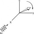
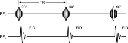
Applying a single RF pulse and producing an image from the resulting FID signal is the simplest of pulse sequences. The saturation recovery pulse sequence was the first of these one-pulse sequences. It uses 90° RF pulses and is sometimes indicated as 90°-90°-90° … (Figure 5-1). An FID is produced after each 90° RF pulse.
 When Mz = 0 and Mxy = M0, the spins are said to be saturated.
In MRI, except for some fast imaging techniques, the data from one pulse sequence are not sufficient to generate an image—only a single line’s worth of data. Rather, the sequence must be repeated many times. It is common for a sequence to be repeated 256 times to obtain data for a 256 × 256 image.
These repetitions are usually spaced by a sizable delay. The time from the start of one pulse sequence to the start of the next pulse sequence is the repetition time (TR).
If the TR is long, on the order of several seconds, the amplitude of the second and successive FIDs equals the amplitude of the first FID. If the TR is short, MZ will not have relaxed to equilibrium, M0. The spins remain partially saturated, resulting in a smaller-intensity FID for the second and successive FIDs. This pulse sequence is a partial saturation pulse sequence.
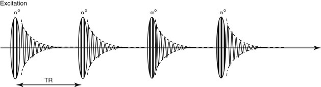
In order to image more quickly, the flip angle can be reduced to α <90°. Using this α-pulse, only part of the longitudinal magnetization, MZ, is flipped into the XY plane to produce signal. This means that there is still some MZ magnetization remaining to be used shortly after the first signal is acquired.
In this manner good images can be made using even short TR values, which allows an increase in imaging speed. The FID signal produced using the lower α flip angle RF pulses creates a smaller signal. However, even with α as low as 10°, a sufficient signal is produced to provide quality images. Therefore in order to use this partial flip angle pulsing strategy the equilibrium MZ magnetization must be large at the beginning of the sequence.
This partial flip pulse sequence is indicated as α°-α°-α° … . The partial flip pulse sequence is shown in Figure 5-2 without the gradient magnetic fields.
Spin Echo and Multi-Echo Spin Echo
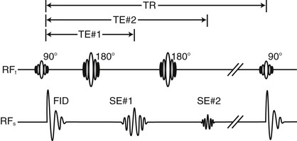
MRI uses the FID for very fast techniques; however, these do not always produce the most useful images. If a 180° RF pulse follows the 90° RF pulse at some later time, an echo signal of the FID can be generated.
This echo signal, called a spin echo (SE), does not follow the 180° RF pulse immediately. The spin echo sequence is the most commonly used two-pulse MRI sequence (see Chapter 6 for further discussion).
 The time between the initial 90° RF pulse and the SE is called time-to-echo (TE).
The time between the 180° RF pulse and the SE equals the time between the 90° and the 180° RF pulses. By controlling the time for the 180° RF pulse, the MRI technologist can control TE. The SE pulse sequence is indicated as 90°-180° … 90°-180° … .
If another 180° RF pulse follows the SE, an additional SE can be generated with a TE that once again is measured from the initial 90° pulse (Figure 5-3). This pulse sequence is a multi-echo spin echo (MESE) pulse sequence. In the three-pulse version of MESE each spin echo, acquired at a unique echo time, TE, is used to create an image with special image contrast characteristics.