(1)
Neurointerventional Radiology Section, Department of Radiology, NYU Langone Medical Center, New York, NY, USA
Abstract
The purpose of this chapter is to summarize the spinal vascular anatomy. After a detailed description of the segmental anatomy, the spinal cord supply will ensue with description of the radiculomedullary arteries, the anterior spinal artery, and the spinal veins. Part of the text, the concept and the figures in this chapter are used with permission from Maksim Shapiro, from the website www.neuroangio.org.
Keywords
Artery of adamkiewiczArtery of lazorthesRadiculomedullarySpinal vasculatureIntroduction
The purpose of this chapter is to summarize the spinal vascular anatomy. After a detailed description of the segmental anatomy, the spinal cord supply will ensue with description of the radiculomedullary arteries, the anterior spinal artery, and the spinal veins. Part of the text, the concept and the figures in this chapter are used with permission from Maksim Shapiro, from the website www.neuroangio.org.
The neural tube/spinal cord can be conceptualized as a cylindrical mass of tissue. For a short period of time after closure of the neural tube, nutritional support can be adequately provided by simple diffusion. When limits of diffusion are exceeded, dedicated vascular supply to the spinal cord and adjacent tissues is established via segmental (metameric) vessels arising from the dorsal aorta. The human embryo is in fact subdivided into 31 somites, each corresponding to a developing metameric segment, that ultimately gives rise to all endo-, ecto-, and mesodermal derivatives. Each somite is supplied by those paired segmental arteries originating from the dorsal aorta. Each segmental vessel supports its endodermal, ectodermal, and mesodermal tissue. Longitudinal anastomoses are then established between segmental vessels, thereby giving rise to new vessels extending craniocaudally along the length of the cord. A grid-like pattern is hence established which is beginning to resemble mature arrangement.
But, as longitudinal vessels develop, hemodynamic need for having a segmental radiculomedullary vessel at each level is diminished. Most segmental vessels diminish to supply only the region of segmental nerve root. The cord is now supplied by the “anterior spinal” artery ventrally. A loose network of arteries with two roughly dominant channels forms the posterior spinal arteries. Finally, only several segmental vessels remain to supply the cord. These are the radiculomedullary vessels (feeding nerve root and cord) of the adult. Other segmental vessels persist as purely radicular (supplying only nerve roots).
The successive transversely oriented segmental arteries anastomose through a number of paraspinal vessels longitudinally arranged along the axis of the spine. These longitudinal channels include the prevertebral artery situated adjacent to the anterolateral aspect of the vertebral body, the pre-transverse anastomoses anterior to the transverse process, as well as the post-transverse spinal anastomotic arcade which extends craniocaudally along both sides of the spinal processes. In the lower lumbosacral spine, a homologous arrangement is recognizable – with the median sacral artery representing the continuation of the aorta and internal iliac arteries considered the homologues of the paravertebral longitudinal arcades, respectively – supplying the corresponding lower lumbar and sacral metamers.
The same segmental arrangement is present in the upper thoracic and cervical spine: the ascending cervical artery which corresponds to the prevertebral artery, the vertebral artery (coursing in the cervical osseous homologue of the transverse process) which is homologous with the pre-transverse anastomotic artery, and the deep cervical artery with the post-transverse anastomosis.
As a consequence of this vascular disposition, the contributions to the anterior spinal axis can originate from any of the above described longitudinal vessels, although most commonly, the dominant cervical radiculomedullary branch (artery of Lazorthes) originates from the lower vertebral artery. The upper thoracic spine is a transitional zone where segmental arteries are supplied by the supreme intercostal artery, which corresponds to the continuation of the pre-transverse anastomosis. As the cord continues to enlarge, perforator or sulco-comissural branches emerge, which unless pathologically enlarged, they are below limits of angiographic resolution.
Arteries
The following discussion is visually recapitulated in Fig. 1, from the website www.neuroangio.org. Throughout the following description, all uppercase letters given in parentheses refer to legends in Fig. 1.
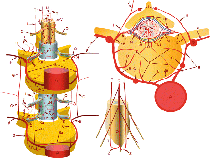
Fig. 1


Schematics of the arterial supply to spinal cord (Courtesy of Dr. Maksim Shapiro from his website, with permission, www.neuroangio.org). A – aorta; B – segmental artery; Ba – intersegmental arterial anastomosis; C – prevertebral anastomotic network; D – direct vertebral body feeding arteries; E – dorsal spinal artery; F – intercostal/muscular artery; G – pretransverse anastomotic network; H – dorsal division of the dorsal spinal artery; I – post-transverse anastomotic network; J – muscular branches of the post-transverse anastomotic network; K – ventral division of the dorsal spinal artery; Ka – radicular artery; La – ventral epidural arcade; Lb – dorsal epidural arcade; M – nerve root sleeve dural branch of the ventral division dorsal spinal artery; N – dural branch of the ventral division dorsal spinal artery; O – radiculopial artery; P – radiculomedullary artery; Q – anterior spinal artery; R – mesh-like pial arterial network; S, T – posterior spinal artery; U, V – pial arterial network (a.k.a. vasocorona) anastomoses between anterior and posterior spinal arterial systems, W – sulcocommissural artery, X – rami perforantes of the peripheral (centripetal) system, Y – central (centrifugal) system of sulcal arteries, originating from pial network of the cord; altogether, the pial network and rami perforantes (R+Y) are called the vasocorona or corona vasorum; Z – rami cruciantes (a.k.a. crux vasculosa, a.k.a. rami anastomotici arcuati)
Related posts:


Stay updated, free articles. Join our Telegram channel
Full access? Get Clinical Tree
