Development of rim-enhancing fluid collection: Splenic abscess
TOP DIFFERENTIAL DIAGNOSES
• Splenic laceration
• Splenic cyst or abscess
• Heterogeneous arterial phase enhancement of spleen
• Splenic tumors
CLINICAL ISSUES
• Many different causes, but 2 most common are
Hematologic disease or hematologic malignancies (sickle cell, myelofibrosis, leukemia, etc.)
Embolic conditions (septic emboli, cardiac emboli from atrial fibrillation, etc.)
• Most cases require no treatment, but rarely surgery or intervention for pain or complications
(Left) Axial CECT in a sickle cell patient demonstrates an enlarged spleen with multiple wedge-shaped acute splenic infarcts . While sickle cell patients can develop a small, calcified autoinfarcted spleen, the spleen may be enlarged in the early stages of the disease.
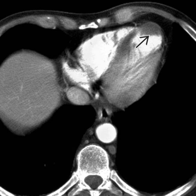
(Right) Axial CECT demonstrates a large, global infarct of the spleen with only a tiny amount of enhancing splenic tissue . Notice the peripheral enhancement (rim sign) at the margins of the infarct as a result of preserved flow through capsular vessels.
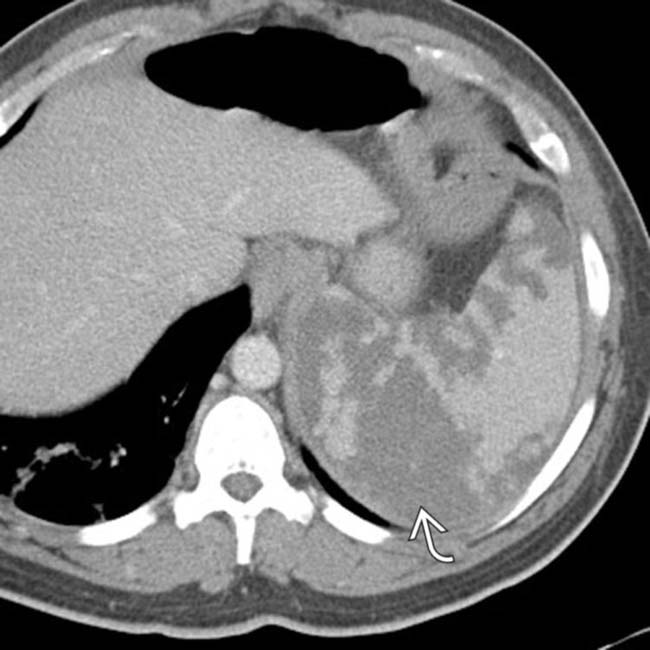
(Left) Axial CECT in a 67-year-old man with a 10-year history of atrial fibrillation, now presenting with acute LUQ pain, demonstrates a peripheral, low-attenuation splenic infarct with straight margins .
(Right) Axial CECT in the same patient identifies a left ventricular thrombus as the source of the arterial embolus to the spleen. Embolic disease is likely the most common cause of splenic infarcts in older patients.
TERMINOLOGY
Definitions
• Global or segmental parenchymal splenic ischemia and necrosis caused by vascular occlusion
IMAGING
General Features
• Best diagnostic clue
Peripheral, wedge-shaped, nonenhancing areas within splenic parenchyma on CECT in patients with LUQ pain
• Location
Entire spleen may be infarcted or more commonly segmental areas
• Size
Variable: Global or segmental
Spleen may or may not demonstrate splenomegaly
• Morphology
Most commonly wedge-shaped areas of nonenhancement when infarct is segmental
– Straight margins indicate vascular etiology (rather than a mass or fluid collection)
– May very rarely be rounded (atypical appearance)
Radiographic Findings
• Radiography
May be associated with lower left lobe atelectasis and pleural effusion on chest x-ray
CT Findings
• NECT
Infarcts may be difficult (or impossible) to visualize without intravenous contrast
Areas of hemorrhagic transformation within infarcts appear hyperdense on NECT
• CECT
Acute findings
– Diagnosis best made on portal venous phase images: Heterogeneous enhancement during arterial phase (due to differential enhancement of red and white pulp) makes identification of subtle infarcts difficult
– Global: Complete nonenhancement of spleen
± cortical rim sign: Preserved enhancement of peripheral rim of spleen in massive infarction due to preserved flow from capsular vessels
Mottled higher density areas within infarcted spleen may represent either tiny islands of residual enhancing splenic tissue or hemorrhage
– Segmental: Wedge-shaped or rounded low-attenuation area usually at periphery of spleen
Can be multiple, especially when caused by emboli
In some instances, accessory spleens (splenules) may be infarcted
Spleen may or may not be enlarged in acute phase
– Complications (< 20% of patients)
Presence of fluid or hematoma surrounding spleen in setting of infarct suggests splenic rupture (most often in setting of large or global infarct)
Development of discrete rim-enhancing fluid collection ± internal gas should raise concern for splenic abscess
Chronic findings
– Infarcts should evolve over time, leaving areas of scarring and volume loss in spleen
Sites of old infarcts may show calcification
Remaining spleen may undergo compensatory hypertrophy
– Multiple repetitive infarcts in sickle cell disease can lead to a small, calcified spleen (autoinfarcted spleen)
– Infarct can develop into splenic cyst (secondary or acquired cyst)
MR Findings
• T1WI
Low signal within area of infarct (can show high T1WI signal due to hemorrhagic infarct)
• T2WI
Heterogeneous high signal within area of infarct
• T1WI C+
Wedge-shaped area of hypoenhancement
Ultrasonographic Findings
• Grayscale ultrasound
Wedge-shaped hypoechoic area(s) within periphery of spleen
– May rarely be rounded or irregularly shaped at center of spleen (atypical)
Bright band sign: Highly echogenic linear bands in area of infarct may be specific sign of infarction
• Color Doppler
Diminished or absent flow in areas of infarction
Angiographic Findings
• Conventional angiography: Main splenic artery occlusion or segmental emboli
Only gold members can continue reading. Log In or Register to continue


 Multiple repetitive infarcts in sickle cell disease can lead to small, calcified spleen (autoinfarcted spleen)


 . While sickle cell patients can develop a small, calcified autoinfarcted spleen, the spleen may be enlarged in the early stages of the disease.
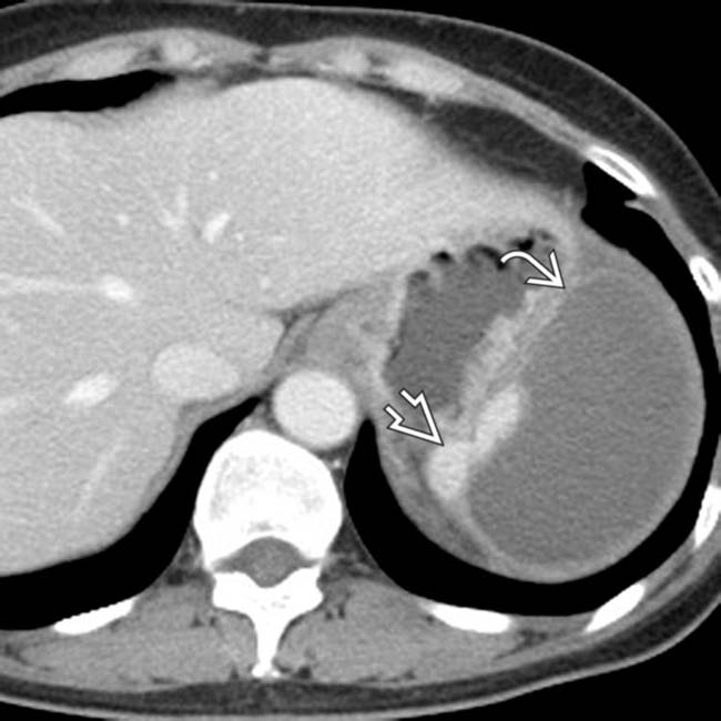
 . Notice the peripheral enhancement (rim sign)
  at the margins of the infarct as a result of preserved flow through capsular vessels.
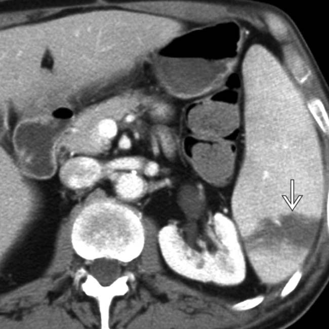
 .
.
 as the source of the arterial embolus to the spleen. Embolic disease is likely the most common cause of splenic infarcts in older patients.


 Acute findings
 ± cortical rim sign: Preserved enhancement of peripheral rim of spleen in massive infarction due to preserved flow from capsular vessels


