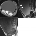
Fig. 1
Serous cystadenoma in a 26-year-old woman. (a) Sagittal T2-weigthed MR image shows a large unilocular cyst. There is a tiny papillary excrescence in the anterior wall of the cyst (arrow). (b) Sagittal T1-weighted MR image shows watery low intensity of the cyst content
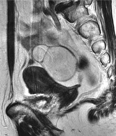
Fig. 2
Serous cystadenoma in a 59-year-old woman. Sagittal T2-weighted image shows bilocular cystic mass posterior to the uterus
Cystadenofibromas typically manifest as a complex cystic mass associated with solid components. On MR imaging, the solid component typically shows distinct low intensity on T2-weighted images, reflecting the fibrous stroma with dense collagen (Fig. 3) [6, 7]. The solid components may contain small or tiny cystic locules [8]. Although serous cystadenomas simulate malignant tumor because of solid components in the cystic mass, their distinct low intensity is an important MR finding for suggesting benignancy. The differential diagnosis of solid mass of low intensity associated with cyst formation includes Brenner tumor and fibrothecomas; both are virtually benign tumors.
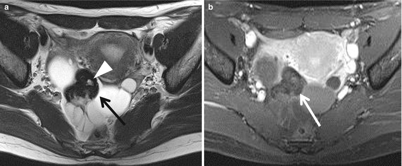
Fig. 3
Serous cystadenofibroma in a 48-year-old woman. (a) Axial T2-weighted MR image shows a complex cystic mass in the right adnexa. The mass contains an irregular-shaped solid component (black arrow) of distinct low intensity in its central portion, which also contains tiny cystic locule (arrowhead). (b) Post-contrast T1-weighted image shows poor enhancement in the solid component (white arrow)
Benign Mucinous Tumors
Mucinous cystadenomas comprise 13 % of benign ovarian epithelial neoplasms, and mean patient age is about 50 years [5]. In contrast to serous cystadenomas, they are bilateral in only 2–3 % of cases. Mucinous cystadenomas are typically multilocular cystic tumors in gross appearance and tend to larger than serous cystadenomas. The cysts usually contain thick, mucinous material, but occasionally contain watery liquid. The locules in mucinous cystadenomas are usually small and multiple. The epithelium in mucinous cystadenoma is characterized by a single layer of tall columnar cells with clear cytoplasm. Histologically, mucinous tumors are further subclassified into intestinal type and endocervical (müllerian) type, and the former comprises over 80 % of mucinous cystadenomas. Up to 10 % of mucinous cystadenomas contain focal component of Brenner tumor.
On imaging studies, mucinous cystic tumor typically shows a characteristic multilocular appearance with varying signals in the locules (“stained glass appearance”) on both T1- and T2-weighted images (Fig. 4) [7]. The locules containing watery fluid usually exhibit low signal on T1-weighted image and high signal on T2-weighted image, whereas those with thick mucinous fluid tend to show increased signal on T1-weighted images. The septi in the cysts exhibit low intensity on T2-weighted images and become enhanced when post-contrast T1-weighted is obtained. The differentiation of benign mucinous cystadenomas and borderline tumors is quite difficult and should depend on postoperative histologic findings. In general, the presence of a thick wall or septa may suggest borderline lesions, while the presence of solid components suggests carcinoma [9]. When the fluid in the locule is very viscose, the signal of the locule may be distinctively low on T2-weighted images, simulating thickened septi or solid mass. In such cases, lack of enhancement on post-contrast images in the area of increased intensity on unenhanced T1-weighted images will be of help for differentiating mucinous locule from solid mass. When mucinous cystadenoma is accompanied by focal component Brenner tumor, it shows distinct low intensity on T2-weighted images and enhanced on post-contrast study (Fig. 5). The presence of calcification on plain CT is also suggestive of Brenner tumor. When multilocular cystic tumors are observed in bilateral ovaries, metastatic ovarian tumors, especially from the colon and appendix, should be considered [10]. Metastatic tumors from mucin-producing carcinoma can show similar morphologic appearance to those of primary mucinous cystic tumor both grossly and microscopically. They commonly involve bilateral ovaries, whereas primary ovarian mucinous tumors are rarely bilateral.
 Ovarian Tumors: Computed Tomography and Magnetic Resonance
 Tumours of the Ovaries: Computed Tomography and Magnetic Resonance
 Cord-Stromal Tumors: Computed Tomography and Magnetic Resonance
 and MR of Benign Ovarian Germ Cell Tumours
 Ovarian Tumors (Serous/Mucinous/Endometrioid/Clear Cell Carcinoma): Clinical Setting and Ultrasound Appearance
 Histopathology


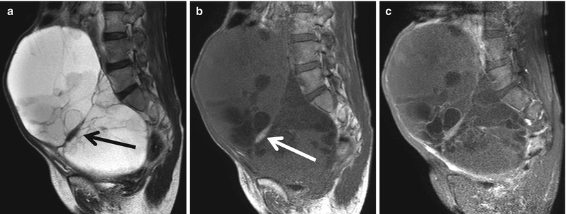
Fig. 4
Mucinous cystadenoma in a 60-year-old woman. (a) Sagittal T2-weighted image shows a large multilocular cystic mass. There is a focal area of distinct low intensity (black arrow), simulating the thickened septum. (b) T1-weighted image shows variable signal intensity in multiple locules. The area of low intensity on T2-weighted image exhibits high intensity (white arrow), suggesting densely viscose fluid. (c) Post-contrast T1-weighted image with fat suppression shows enhancement in the septi
Related posts:
Ovarian Tumors: Computed Tomography and Magnetic Resonance
Tumours of the Ovaries: Computed Tomography and Magnetic Resonance
Cord-Stromal Tumors: Computed Tomography and Magnetic Resonance
and MR of Benign Ovarian Germ Cell Tumours
Ovarian Tumors (Serous/Mucinous/Endometrioid/Clear Cell Carcinoma): Clinical Setting and Ultrasound Appearance
Histopathology
Stay updated, free articles. Join our Telegram channel
Full access? Get Clinical Tree
